Download
1 / 39
390 likes | 407 Vues
Explore the pathophysiology, symptoms, diagnosis, and treatment of respiratory conditions including respiratory failure, pleural effusions, lung cancer, bronchiectasis, and occupational lung disease. Understand the causes, symptoms, and diagnostic methods for these conditions to provide effective care.

E N D
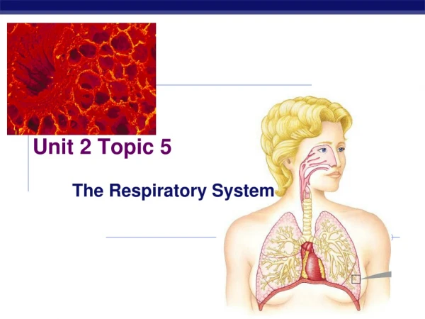
Respiratory diseases nur124 – session 5 Nadeeka Jayasinghe
OBJECTIVES Discuss pathophysiology, symptoms, diagnosis, treatment of: • Respiratory Failure • Pleural Effusions • Lung Cancer • Bronchiectasis • Occupational Lung Disease • Traumatic Disorders Of The Lung
RESPIRATORY FAILURE • A process where the respiratory system fails in one or both of its gas exchange functions 1. Oxygenation 2. Carbondioxide elimination • Hypoxic(Type 1) or Hypercapneic (Type 2)
HYPOXIX RESP FAILURE (TYPE 1) • Arterial oxygen tension (PaO2) <60mmHg with low or normal arterial carbon dioxide tension (PaCO2). • Most common form of respiratory failure • Can be associated with most lung diseases which involve fluid overload or alveloar collapse • Examples : Cardiogenic/non-cardiogenic pulmonary edema, pneumonia
HYPERCAPNEIC RESP FAILURE • Arterial carbondioxide tension (PCO2) higher than 50mmHg. (Hypercapniea) • Can lead to hypoxemia if they are breathing in room air • Blood pH will be less than 7.35 • Can develop over minutes to hours • The pH levels depend on the bicarbonate levels in the body which in turn is depedant
RESP FAILURE - CAUSES • Abnormalities in any of the components of the respiratory system including the airway, alveoli, central nervous system (CNS), peripheral nervous system, respiratory muscles and chest wall. • Pharmacological, structural and metabolic disorders of the CNS may lead to respiratory depression. This leads to hypoventilation or hypercapnea. (tumors in brain stem, sedatives, overdoses)
RESP FAILURE - CAUSES • Disorders of the peripheral nervous system
RESP FAILURE - DIAGNOSIS • Arterial Blood Gases • Chest xray • ECG (not essential but can rule out possible cardiac causes ) • Pulmonary function tests
PLEURAL EFFUSIONS • WHERE IS THE PLEURAL SPACE OF THE LUNG? • Definition?
PLEURAL EFFUSIONS • The pleura is the thin membrane that lines the surface of the lungs and inside the chest wall outside the wall. • A pleural effusion is an abnormal amount of fluid around the lung. • Normally, 5-10ml of fluid is in the pleural space allowing the lungs to move smoothly during respiration.
CAUSES OF PLEURAL EFFUSIONS • Congestive heart failure • Pneumonia • Liver disease (cirrhosis) • End stage renal disease • Nephrotic syndrome • Cancer • Pulomonary embolism • Lupus and other autoimmune conditions
Why does excessive fluid accumulate? • Due to fluid overload – congestive heart failure, renal and hepatic disease. • Inflammation – pneumonia, autoimmune disease
SYMPTOMS • Shortness of breath • Chest pain upon breathing (pleuritic) • Fever • Cough • Decreased chest movement and breath sounds on affected side • Bronchial breathing
Diagnosis • Auscultation and percussion (very difficult to rule out) • Chest xray – (white space at the base of lungs) • CT scan • Ultrasound – assists with drainage of fluid
TREATMENT – Pleural effusions • Thoracentesis – a needle is inserted into the chest wall between the 6/7/8th intercostal space on mid-axillary line into the pleural space and fluid is drained out. • Pleural tap may be left in for a few hours to drain fluid over time • Fluid can be used to determine – protein content, cell count, infection (via culture), fungus, gram stain, lipids etc.
LUNG CANCER • Pulmonary carcinoma • Small cell carcinoma vs non small cell carcinoma • 80-90% of lung cancers are from long term tobacco smoke exposure • 10%-15% occur in patients who have never smoked (genetics, pollution, asbestos exposure, second hand smoking)
SYMPTOMS • Respiratory : coughing, hemoptysis, wheezing, shortness of breath • Systemic: weight loss, fever, clubbing of the fingernails, fatigue • Symptoms due to the mass pressing on adjecant structures: chest pain, bone pain, superior vena cava obstruction, swallowing difficulty
DIAGNOSIS • CT SCAN • LUNG BIOPSY (via brochoscope and/or CT guided biopsy)
Treatment • Surgery : Pulmonary function tests must first reveal that the patient is well for surgery. Lobectomy, wedge resection and pneumonectomy are options. • Radiotherapy: Can be given with chemotherapy. For patients who are not suitable for surgery • Chemotherapy: Improves survival but has severe side effects.
BRONCHIECTASIS • A disease where the lung is abnormally widened due to mucus blockage • Can develop at any age. But common at birth (congenital bronchiectasis) • Infection - TB, influenza, pneumonia, cystic fibrosis • Due to a blockage in your airway: due to a mass, or an inhalation of a solid (food etc)
BRONCHIECTASIS • Mucus to build up causes bacteria growth and severe infection. Over time, the airways loses it’s ability to ventilate adequately. • Can lead to respiratory failure, heart failure and collapsed lung.
Symptoms • Shortness of breath • Hemoptysis • Wheezing • Chest pain • Fatigue • General lethargy and feeling unwell
Diagnosis • Chest CT scan • Chest xray – will show airway abnormalities • Blood tests – infection, other conditions that may be contributing factors • Lung function tests – capacity of lungs, if breathing volumes are affected • Bronchoscopy – blockages of airway, bleeding etc.
Treatment • Treatment of underlying conditions • Antibiotic therapy • Chest Physiotherapy • Bronchodilators, steroids, oxygen • Surgery
OCCUPATIONAL LUNG DISEASES • Broad group diagnosis • Inhalation of dust, chemical and proteins • “Pneumoconiosis” – diseases associated with inhaling mineral dust • The exposure of different particles result in different diseases Asbestos exposure - Asbestosis Silicon exposure – Silicosis Coal / mineral dust - Pneomoconiosis
OCCUPATIONAL LUNG DISEASE 1. ASBESTOSIS: • Asbestos – used for industrial work – breaks into fibers when shattered • Industrialization exposed large communities to asbestosis but symptoms did not develop till later in life • Asbestosis causes scarring, fibrosis, lung cancer, pleural effusions, plaques. • Dyspnoea
OCCUPATIONAL LUNG DISEASE 2. SILICOSIS: • Develops decades after exposure • Silica nodules in the lungs • Acute (higher mortality) vs chronic silicosis (silicotic nodules develop into lesions) • Silicosis increases risk of TB and immune related diseases (systemic arthritis and SLE) • Associated with increased risk of lung cancer
OCCUPATIONAL LUNG DISEASES 3. PNEMOCONIOSIS (CWP): • Long term exposure to coal dust • Also known as black lung – small spots in upper lungs that reflect coal inhalation • Progresses into fibrosis. Similar to Silicosis – destroys lung architecture • Exposure to coal dust – airflow obstruction, chronic bronchitis, rheumatoid arthritis • Stomach cancer has been associated with coal ingestion
TRAUMATIC DISORDERS OF THE LUNG • PNEUMOTHORAX: • An abnormal collection of air or gas in the pleural space that separates the lung from the chest wall • Spontaneous pneumothorax – occurs without an apparent cause in the absence of lung disease • Secondary pneumothorax – occurs in the presence of significant lung pathology
TRAUMATIC DISORDERS OF THE LUNG • In a minority of cases, the amount of air in the chest increases markedly when a one-way valve is formed by an area of damaged tissue, leading to a tension pneumothorax. This condition is a medical emergency.
Causes (Pneumothorax) • Physical trauma to the chest wall • Blasts • Complication from a medical or surgical intervention • Long term mechanical ventialation
Signs and symptoms • Shortness of breath • Chest pain (mild to severe – depending on stage) • Hypoxia – leads to cyanosis • Hypercapnia – confusion
DIAGNOSIS • Chest xray • CT scan • Auscultation • Observation and assessment of changes in patient condition (tracheal deviation, changes to the shape of chest wall)
Management • Not all pneumothoraces require treatment • Immediate needle decompression (if in an emergency setting) • Chest tube insertion • Pleurodecis (