ANAESTHESIA WITH CONCURRENT RESPIRATORY DISEASES
890 likes | 1.42k Vues
ANAESTHESIA WITH CONCURRENT RESPIRATORY DISEASES MODERTOR BY- DR SUCHIT KHANDUJA DR GIAN CHAUHAN JR ANAESTHESIA. Preoperative Preparation. General assessment- This involves history, examination and investigation .

ANAESTHESIA WITH CONCURRENT RESPIRATORY DISEASES
E N D
Presentation Transcript
ANAESTHESIA WITH CONCURRENT RESPIRATORY DISEASES MODERTOR BY- DR SUCHIT KHANDUJA DR GIAN CHAUHAN JR ANAESTHESIA
Preoperative Preparation General assessment-This involves history, examination and investigation. History. Ask about symptoms of wheeze Cough Sputum production, Haemoptysis Chest pain Exercise tolerance, Orthopnoeaand paroxysmal nocturnal dypsnoea
Diagnosis of chronic chest complaints such as asthma or bronchiectasis is often known. • Present medication and allergies are noted, and a history of smoking sought. • Previous anaesthetic records may be available and can help in planning care.
Examination. Inspect for • Cyanosis • Dyspnoea • Respiratory rate • Asymmetry of chest wall movement • Scars, cough and sputum colour.
Percussion and auscultation of chest may suggest • Areas of collapse and consolidation, • Pleural effusions, • Pulmonary oedema or infection. • Corpulmonale may be evident as peripheral oedema and raised jugular venous pressure • Enlarged lymph nodes in the neck may suggest lung cancer.
Investigations. Leucocytosis may indicate active infection, and polycythaemia chronic hypoxaemia. ABG should be performed in patients who are dyspnoeic with minimal exertion and the results interpreted in relation to PIO2. Preoperative hypoxia or carbon dioxide retention indicates the possibility of postoperative respiratory failure. May require a period of assisted ventilation on the Intensive Care Unit.
Pulmonary function tests, provide baseline pre-operative measurements. • Chest clinic has charts to compare these results against those predicted for the patients age, sex and weight. • The results are also compared against the patient's previous records to assess current disease control.
FEV1.0 (Forced Expiratory Volume in 1 second) and FVC (Forced Vital Capacity) are commonly measured. A reduction in the FEV1.0:FVC ratio indicates obstructive airways disease. (The normal is 0.75 (75%) or more). A reduction in FVC occurs in restrictive lung disease. • An FEV1.0 or FVC less than 70% of predicted, or an FEV1.0:FVC ratio less than 65%, is associated with an increased risk of pulmonary complications.
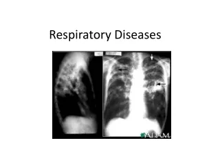
Chest X-rays may confirm effusions, collapse and consolidation, active infection, pulmonary oedema, or the hyperinflated lung fields of emphysema. • ECG may indicate P-pulmonale, a right ventricular strain pattern (dominant R waves in the septal leads) or right bundle branch block.
In patients with poor respiratory function premedication (if used) must not cause respiratory depression. • Opiates and benzodiazepines can both do this • Are best avoided if possible, or used with caution. • Humidified oxygen may be administered . • Anticholinergic drugs (e.g. atropine) may dry airway secretions and may be helpful before ketamine .
Specific Respiratory Problems Coryza (common cold) • Typically, children experience six to eight URIs per year • May be even more frequent among young children attending nursery school or day care • 30%–40% of URIs are caused by rhinoviruses; • Other viruses—including coronavirus, respiratory syncytial virus, and parainfluenza virus—contribute significantly to the etiology of the disease.
Patients may present with undiagnosed infections including croup (laryngotracheobronchitis), influenza, bronchiolitis, herpes simplex, pneumonia, epiglottitis, and strep throat. • Most viral URIs are self-limiting • May produce airway hyperreactivity that persists for several weeks after infection. • Viral invasion of the respiratory mucosa may render the airway sensitive to secretions or potentially irritant anesthetic gases.
Viral invasion of the respiratory mucosa may render the airway sensitive to secretions or potentially irritant anesthetic gases. • Bronchial hyperreactivity resulting from viral infections may be neurally mediated. • Atropine, for example, has been shown to block airway hyperreactivity. • Viral infections increase the response of airway smooth muscle to tachykinins . • De Soto et al. (9) found that children with symptoms of a URI had a significant increase in the risk of postoperative arterial oxygen desaturation and laryngospasm.
Independent risk factors for adverse respiratory events in children with active URIs include Use of an ETT in a child <5 yr old. Prematurity (<37 wk). History of reactive airway disease. Paternal smoking. Surgery involving the airway, Presence of copious secretions. And presence of nasal congestion.
Preoperative Assessment Child is presenting for an emergent procedure • Presence of a URI should be elicited • Will alert the anesthesiologist for complications • Modification of the anesthetic management to reduce any risk.
Children for elective procedures with suspected URI • Require careful preoperative assessment • Detailed history and physical exam to be done. • Lungs should be auscultated to exclude any lower respiratory tract involvement, • A chest radiograph should be considered if the examination is questionable. • Patient should be evaluated for fever, dyspnea, productive cough, sputum production, nasal congestion, lethargy, and wheezing.
Nasal congestion, sputum production, and a history of reactive airway disease were identified as predictors of adverse respiratory events • Confirmation of URI by a parent found to be a better predictor of laryngospasm than symptom criteria . • Children with congenital heart disease can have URI confused with CHF.
Should be able to undergo surgery …. • Children presenting with uncomplicated URI • Who are afebrile with clear secretions and appear otherwise healthy • Those with noninfectious conditions
Should be postponed for surgery…… Children with more severe symptoms—including • Mucopurulent secretions • Productive cough • Fever >38°C • Lethargy • Signs of pulmonary involvement • If a bacterial infection is suspected Their elective surgery postponed for a minimum of 4 wk or more(if bact. Infection is suspected)
INVESTIGATIONS • Analysis of nasopharyngeal swabs or aspirates for viral isolation • Measurement of white blood cells counts • The chest radiograph is also of little utility
Anesthetic Management • Directed at minimizing secretions and avoiding stimulation of a potentially sensitive airway. • Airway be suctioned (under deep anesthesia) to remove excess secretions • Ensure that the patient is adequately hydrated • Humidification may also be important in children with URIs, particularly for long cases.
Anticholinergicsglycopyrrolate or atropine may be useful in reducing secretions and attenuating vagally-mediated hyperreactivity. • Bronchodilator premedication has also been suggested • Preop treatment with corticosteroids and salbutamol minimized intubation-evoked bronchoconstriction more effectively than inhaled salbutamol alone.
Use of an ETT should be avoided • ETT is likely the airway of choice for surgery of the oropharynx and neck, major thoracic and abdominal surgery, and operations lasting more than a couple of hours. • LMA, is a safe alternative for some procedures • Complications in children with mild URIs was similar between sevoflurane and halothane. • Sevoflurane provided a more rapid recovery profile
Can prefer to extubate under deep anesthesia to avoid reflex constriction of the airways. • Also can extubate when the patient is awake, believing that a patient with intact reflexes is in a better position to clear secretions and respond to the tactile stimulation of ETT removal.
Asthma • Asthma causes hyper-responsive airways with oedema, inflammation and narrowing due to smooth muscle spasm. • Characteristically reversible, unlike chronic obstructive pulmonary disease • Elective cases should not be undertaken unless asthma is well controlled • In poorly controlled asthma a short course of steroids is often effective in improving control of the disease. • Patients on preoperative steroids will need extra perioperative supplementation if they are taking more than the equivalent of 10mg of prednisolone a day.
Preoperative assessment • Assessed by the frequency and severity of attacks • Hospital and intensive care admissions seen • Examination may reveal expiratory wheezes, use of accessory muscles or an over-distended chest. • Peak expiratory flow rates (PEFR) pre- and post-bronchodilator should be measured
Blood gas analysis is usually reserved for severe disease (breathlessness on minimal exertion). • Before surgery, patients should be free of wheeze • PEFR greater than 80% of the predicted or personal best value • Severe asthmatics may require their inhalers being changed to nebulisers. • Inhaled steroid dose may have to be increased or oral steroids commenced (Prednisolone 20-40mg daily) one week prior to surgery
Perioperative management • Consider converting inhaled beta 2 agonists such as salbutamol to the nebulised form • Give nebulisedsalbutamol (2.5-5.0mg) with premedication. • Avoid aspirin or NSAIDs and any other allergens known to the patient. • If applicable local or regional anaesthesia used alone will avoid the problems of general anaesthesia. • If general anaesthesia is required, the addition of regional techniques can reduce operative volatile anaesthetic and post operative opioid requirements and the likelihood of respiratory complications.
Ketamine and all the volatile agents are bronchodilators. • Avoid thiopentone • Airway manipulation should be kept to a minimum • Controlled ventilation with the use of neuromuscular blocking drugs will be needed for major or long procedures • Atracurium and tubocurare,high dose opoids should be avoided as they release histamine. • Deriphylline with b2 adrenergic agonist and halothane should be carefully used together. • Capnograph must be carefully noted for signs of bronchospasm.
Residual neuromuscular blockade must be fully reversed • Extubation can occur when spontaneous ventilation is resumed and oxygenation is adequate. • Deep extubationdec. chances of laryngospasm. • Extubation induced laryngospasm can be blunted by lidocaine 1-2 mg/kg prior to extubation.
Postoperative care • Adequate analgesia is vital. • Humidified oxygen is continued for up to 72 hours following major abdominal or thoracic surgery together with regular physiotherapy until the patient regains mobility. • Maintenance of hydration with intravenous fluids is required until oral intake is sufficient. • Usual anti-asthmatic medications are resumed immediately • May require intravenous steroids to temporarily replace oral • Nebulised bronchodilators to replace inhalers if the patient cannot take a deep breath • Failure to ensure adequate postoperative oxygenation and ventilation may require admission to an intensive care area.
Anaesthesia in patients with Chronic Obstructive Pulmonary Disease • Chronic obstructive pulmonary disease (COPD) is a very common respiratory disorder that affects many people across the world. • Long term survival of patients with severe COPD undergoing any type of surgery is poor (47% 2 year mortality) • Significant risk of postoperative morbidity, especially pulmonary complications
Pathophysiology of COPD • Inflammatory disease of the lungs that is characterised by airflow limitation that is not fully reversible • Complicated by significant systemic manifestations and co-morbidities. • Usually secondary to inhaled noxious particles or gases • Most common of which worldwide is cigarette smoke
The lung pathology in COPD is a combination of inflammatory small airways disease(obstructive bronchiolitis) and parenchymal destruction (emphysema). • The small airways disease leads to obstruction and air trapping • The loss of lung parenchyma decreases gas transfer, reduces the pulmonary capillary bed and worsens VQ mismatching. • Reducing the parenchymal support of the walls of the small airways contributes to the airflow limitation.
Causes hypoxaemia and sometimes hypercarbia. • Direct result of COPD can be corpulmonale • Respiratory and skeletal muscle wasting and weight loss can occur • Co-morbid diseases, especially cardiovascular are more prevalent
Clinical Features of COPD • Dyspnoea • Wheeze • Cough with or without sputum production. • Onset of COPD is insidious • Most patients are symptomatic either with cough or progressive dyspnoea long before they present to medical services.
Diagnosis of COPD should be considered in all patients over forty years old with a significant smoking history (>10 pack years) with some symptoms. • Mainstay of treatment of COPD is bronchodilation both for maintenance and for exacerbations. • β-agonists and anticholinergics (ipratropium bromide and tiotropium bromide) are used; the latter are proposed to have an additional effect of relieving air trapping.
Long term inhaled steroids are usually only indicated in patients with severe COPD and repeated exacerbations or who have co-existent asthma. • Oral steroids are beneficial in the treatment of exacerbations.
Preoperative assessment History and examination • Establish exercise tolerance, particularly hills and stairs • Enquire about frequency of exacerbations, hospital admissions and previous requirements for invasive and non-invasive ventilation. • A smoking history is vital
Cough and particularly sputum production has been shown to be an independent risk factor for postoperative pulmonary complications in COPD. • Clear history regarding co-morbid conditions is vital. • Symptoms and signs of active infection should be sought including green or purulent sputum, increased dyspnoea, wheeze and signs of consolidation. • Nutritional status should be assessed • Patients with both high and low BMI have increased risk.
Investigations • Chest X-ray is useful to exclude active infection and occult malignancy • The presence of extensive bullous disease highlights the potential risk of pneumothorax. • ECG may reveal right heart disease (right ventricular hypertrophy or strain). • Spirometry is used to clarify diagnosis and assess severity
GOLD Classification of COPD (based on post bronchodilator FEV1) • Stage I: Mild FEV1/FVC <0.70 • FEV1 ≥ 80% predicted • Stage II: Moderate FEV1/FVC < 0.70 • FEV1 50 - 80% predicted • Stage III : Severe FEV1/FVC <0.70 • FEV1 30 - 50% predicted • Stage IV: Very Severe FEV1/FVC 0.70 • FEV1 <30% predicted or FEV1 < 50% predicted and chronic respiratory failure
Simple exercise tests such as stair climbing and the 6 minute walk test are safe and simple to perform • Arterial blood gas measurement is useful: PaCO2 > 5.9 kPa and PaO2 <7.9 kPa (on room air) are both associated with a worse prognosis.
Preoperative optimisation • • Smoking cessation • Current smokers are at far greater risk of developing postoperative pulmonary complications. • should be stopped at least eight weeks before surgery in order to obtain maximum benefit
Optimal drug treatment. • Almost all patients with COPD benefit from at least one dose of nebulised bronchodilator preoperatively • High doses of nebulised β-agonists can cause or exacerbate tachyarryhthmias and cause hypokalaemia. Nebulisedanticholinergics can increase sputum viscosity
Treatment of infection/ exacerbation • Current infection or exacerbations are a contraindication to anaesthesia. • Should be treated with both β-agonist andanticholinergic therapy, preferably in nebulised form, and a short course of systemic steroids. • Prophylactic antibiotics over the operative course may be beneficial
Physiotherapy • Preoperative physiotherapy is important in sputum producing COPD patients to clear any retained sputum that may cause intraoperative bronchial plugging or pneumonitis. • Pulmonary rehabilitation, may also have a role
Principles of perioperativeanaesthetic management Airway and ventilation • Bronchospasm may occur on induction, during airway instrumentation and on extubation. • May need aggressive administration of bronchodilators to prevent or treat acute hypoxaemia and hypercarbia • Avoidance of endotracheal intubation, if possible, may reduce the risk of bronchospasm. • Extubation should be carried out with the patient awake and sitting up.
V/Q mismatch increases under general anaesthesia and in the supine position • Combined with atelectasis, this leads to worsening hypoxaemia • Pronounced if the patient is obese or is in the Trendelenburg or lithotomy position. • Supplemental oxygen and positive pressure ventilation may be necessary.