Download
1 / 35
390 likes | 524 Vues
This program aims to improve lung function, gas exchange, and ventilation in respiratory diseases through targeted exercises and therapies. It addresses bronchial obstruction, inflammation, and ventilation issues, enhancing overall respiratory health. Contraindications for specific exercises are highlighted, and objectives for bronchitis rehabilitation are outlined. Techniques such as drainage exercise, postural drainage, and massage are emphasized for effective treatment. The program aims to stabilize irreversible lung changes, restore respiratory function, and enhance cardiovascular health and psychological well-being. Various clinical manifestations of respiratory diseases, including dyspnea, cough, and chest pain, are discussed, emphasizing the importance of tailored rehabilitation strategies.

E N D
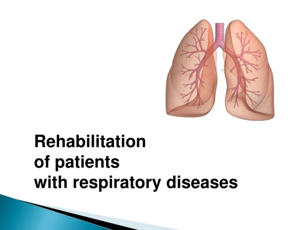
Rehabilitation of patients with respiratory diseases
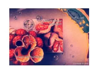
When pulmonary disease in the organismthere is a violation of respiratory function. This is due to deterioration of the elasticity of the lung tissue. • Also disrupted the normal gas exchange between blood and alveolar air. • Bronchial conductivity decreases as a result of spasm of the bronchi, thickening of their walls, mechanical blockage that occurs with increased sputum,
THE OBJECTIVES AND PRINCIPLES OF REHABILITATION OF PATIENTS WITH PULMONARYDISEASES Special problems of rehabilitation include: • elimination of the inflammatory focus; • improvement of bronchial obstruction; • increasing of lung ventilation; • elimination of inconsistency between alveolar ventilation and pulmonary blood flow; • improving the lung drainage; • economization of the respiratory muscles’ work by increasing their capacity and cooperation. The overall objectives of rehabilitation are: • achieving the regression of reversible and stabilization of irreversible changes in the lungs; • restoration and improvement of respiratory function and cardio-vascular system, the psychological status and capability.
Contraindications for practicing exercise therapy in case of respiratory diseases • Respiratory insufficiency of the third degree • Presence of lung abscess breaking through in the bronchi • Coughing up blood, or threat of it • Asthmatic status • Full pulmonary atelectasis • Accumulation of large amounts of fluid in the pleural cavity
Objectives of physical rehabilitation for bronchitis: • 1) Improving blood circulation and lymph flow; • 2) Decrease or elimination of inflammatory changes in the bronchi; • 3) Restoration of the drainage function of bronchi; • 4) Prevention of chronic bronchitis, pneumonia; • 5) Increasing local and general resistibility of the bronchial tree and cold resistance of the body.
Objectives of physical rehabilitation for bronchitis: • In case of obstructive syndrome is advisable to use sound exercise, followed by breathing exercises. After 2-3 weeks of sound exercise inspiration and exhalwith resistance should be included (breath alternately with different corner of the mouth, different nostril.) • In case of purulent chronic bronchitis the thing of high importance is drainage exercise and postural drainage which are carried out regularly (2-4 times a week). They contribute to a more complete removal of purulent sputum from the bronchi
Application of massage on the chest is very significant. It contributes to a better allocation of phlegm and facilitates breathing • These activities will contribute to: • "purification" of bronchi of purulent sputum • improvement of their drainage function • normalization of breathing • to mobilize compensatory ventilation mechanisms
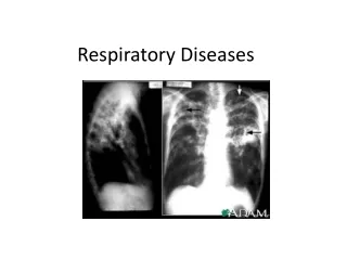
The main clinical manifestations of respiratory diseases 1. Changing the rate and rhythm of breathing - rapid breathing as a compensatory adaptation: decreasing the respiratory surface, in feverish conditions (high temperature), with sharp pains that do not allow to breathe deeply
Dyspnea • - difficult breathing disorder, feels like a lack of air.Рatient breathes deeply and often. • There are three types of dyspnea: 1) inspiratory (difficult inhalation)- is more common in the upper airway constrictions; 2) expiratory (exhalation difficult) - is observed with decreasing elasticity of the lung tissue and narrowing of small airways, • and 3) mixed - hampered both phases of respiration. Breathing usually quickens. Occurs in many diseases of the lungs. Strong degree of dyspnea in which the patient is choking, suffocation is called. Choking, there are attacks, called asthma.
Cough -a reflex act • Reason for the cough - irritation of the respiratory tract foreign bodies or abnormal products (sputum, blood). Cough frees airways of these products. Distinguish between dry cough without sputum and wet with sputum.
Dyspnea • is the main clinical feature of pulmonary failure, and • its functional feature - the inability of the respiratory system to ensure a particular exercise. Typically, when respiratory failure show signs of dysfunction of the cardiovascular system, because cardiovascular failurejoinstorespiratory failure
Pain in the chest • - this sharp stabbing pains or weaker and longer; • characterized their relation with breathing (pain when taking a deep breath, when you cough). • Pain are more often in the side. • Apart from these major manifestations may also be: • - feeling hot,, fatigue, etc., • - sometimes chills, • - fatigue, • - headaches etc.
In the clinical picture of acute pneumonia syndromes are the following: • Intoxication - general weakness, pallor, loss of appetite; • General inflammatory changes - fever, chills, a sharp rise in temperature to 39-40 ° C, leukocytosis, increased erythrocyte sedimentation rate (ESR); • Inflammatory changes in the lung tissue - the appearance of cough and sputum, change in the frequency and nature of respiration, the emergence of moist rales; • Involvement of other organs and systems - changes in the cardiovascular, nervous and other systems.
The main objectives of physical rehabilitation (exercise therapy) for pneumonia: • improving blood circulation, lymph circulation in the lungs to accelerate the resorption of inflammatory exudate and prevent complications; • to contribute to a more intensive removal of sputum • to strengthen the respiratory muscles, the increase of lung excursion • normalization of respiratory mechanics: slowing and deepening of breath • restoration of adaptation to physical stress • rehabilitative effect on the cardiovascular , respiratory, nervous and other systems of the body to improve their function and maintain general physical performance.
Exercise therapy for pneumonia: • During the period of bed rest, from 3d-5th day at lying position and sitting on a bed, legs dangling, dynamic exercises for small and medium-sized muscle groups are applied, as far as static and dynamic breathing exercises. • The ratio of general developmental and breathing exercises - 1:1, 1:2, 1:3. • There should be no increased heart rate by more than 5-10 blow/min. • Exercises have to be performed in a slow and mid-tempo, each one repeated 4-8 times with a maximum range of motion. • Duration of treatment - 10-15 min, individual classes - 10 min 3 times a day.
On ambulation regime or bed rest mode • from 5th - 7th day in sitting or standing positions bed rest exercises are continued to be used, however the dosage is increased, including exercises with objects for the major muscle groups. • The ratio of respiratory exercises and general exercises -1:1, 1:2. • Pulse increase is allowed up to 10-15 blow/min , the number of repetitions of each exercise is increased up to 8-10 times the average rate. • Estimated exercise time is 15-30 minutes, also walking is used. • Exercises are performed individually. • The total duration of exercise during the day is up to 2 hours, the classes сan be individual, few-group and independent .
From the 7th-10th day • (not earlier) patients are transferred to the general regime. • Therapeutic exercises are similar to those used on ambulation regime, but with a greater load, causing increased heart rate - up to 100 b/min. • The duration of one exercise set is 40 minutes, application ofexercises, walking, training on simulators andgames takes about 2.5 hours per day.
Lung Emphysema Structure of the lung in normal and emphysema • Lung emphysema is characterized by persistent expansion of the lungs, increasing their airiness due to lower elasticity of lung tissue, trophic changes of the alveoli and their magnification. • As an independent form the disease is rare, often being a consequence of chronic lung diseases such as chronic pneumonia, chronic bronchitis, bronchial asthma, pulmonary tuberculosis. normal emphysema
Other causes of pulmonary emphysema • may be permanent mechanical distension of lungs in case of heavy physical work associated with straining and thus air delay, as well as age-related changes , that is why emphysema is often observed in the elderly. • Due to the increase of lung volume and low mobility of the diaphragm respiratory excursion of the chest is reduced. • The patient has shortness of breath, initially expiratory type,isdifficult to breathe, and later, when heart inspiratory develops, mixed failures are developing. • The chest becomes barrel-shaped.
Exercise therapy task with emphysema The objectives of the physical exercise with emphysema include: • Learning proper breathing, where there is a deeper breath • Reducing the stress of respiratory muscles, strengthening them • Improving the security of blood oxygen • Increasing mobility of the chest • To promote drainage of bronchial and lung cavity • Improving physical performance. • There are also contraindications, they are similar to the contraindications with other respiratory diseases, but at the presence of "rusty" sputum exercises can be applied. • Extended breath reduces the amount of residual air and thus improves gas exchange.
Exercise therapy in the first period of disease development • Exercise therapy is used in non-acute phase, in the absence of severe cardiovascular disease. • The systematic application of exercise in the first period of the disease is capable of maintaining sufficient elasticity of the lungs and chest mobility. • Activating the mechanisms of hemodynamic support and strengthening the heart muscle, exercise contribute to the maintenance of compensation of the cardiovascular system. Use of low and moderate exercise intensity. • Exercises of high-speed nature have the restrictions applied with involvement in the movement of small muscle groups. Grunting and breath holding are not allowed. • Great attention should be paid to the exercises, providing the mobility of the chest and diaphragm breathing exercise. Exercises are necessary to be alternated with breathing and pauses for rest .
Exercise therapy in the second period of disease development • In the second period of the disease exercise therapy (considering the changes in the lung tissue) primarily contributes to the formation of compensation that improves lung ventilation and increases gas exchange in lungs. • This is achieved through exercises designed to strengthen the muscles involved in exhalation, increasing mobility of the chest, the development of diaphragmatic breathing, learning mechanism of breathing with prolonged exhalation. • Prolonged exhalation reduces the amount of residual air and improves gas exchange. The increase in the mobility of the chest and diaphragm excursion creates conditions which facilitate the work of the heart. • Exercises of tilting and rotating the torso are widely used.
Exercise therapy in the second period of disease development • In the third period of the disease exercise therapy is used with moderate stagnation concerningbed rest. • Exercises with a small load (movement in the distal extremities in slow and medium tempo) • in a reclining position with a raised trunk should provide improved peripheral circulation and dosed venous flow to the heart (rhythmic movements of the limbs with incomplete amplitude at a slow tempo). • They should be alternated with pauses for relaxation and breathing exercises with extended expiration. Diaphragmatic breathing with retraction of the abdominal wall on the exhale is activated. • Then movements are combined with in-depth breathing. • Part of the exercise is completed by compression of the chest as you exhale (performed by instructor). • Muscle relaxation exercises are widely used. • The density of exercises is low.
With a significant reduction of heart failures • it is possible to begin doing the exercises in sitting and lying down positions. • Exercises with the participation of the large muscle groups, initially with a small number of repetitions (2-4 times). • With the gradual increase of the amplitude of movement. • Exercises that help to enhance the mobility of the chest are performed smoothly, at a slow tempo. • It is necessary to combine these movements with the breath, often including a pause for the rest.
The ambulation regime • While improving the condition of patients and improving their adaptability to physical stress the ambulation regime has to be assigned. • The number of movements of the large muscle groups is increased, exercises are performed in lying, sitting and standing positions. walking at a slow tempo (60-70 steps per minute) in combination with an extended expiration are also performed. • Distance is gradually increased up to 200-300 meters
The free regime • In case of satisfactory adaptation to the used loads the patients are to be transferred to the free regime. • Load at physiotherapy exercises becomes larger (mainly due to the increased range of motion and the number of repetitions). • Walk distance is increased up to 400-500 meters, walking up the stairs in the range of 2-3 floors is introduced.
Training of diaphragmatic breathing • In cases of irreversible changes of the lung tissue and developing fibrosis, gymnastic exercises should be directed to the formation of the compensations which improve ventilation and increase gas exchange. • Diaphragmatic breathing and breathing exercises are applied in this case. Exercises with a longer exhalation are performed(exercise with pronunciation of the sounds during the exhale). • Diaphragmatic breathing training in lying and standing positions. • The patient stands with legs wide apart, moving hands sideways, takes a breath, and then, turning hands forward and leaning down, produces a slow exhale during which the abdominal muscles are drawn in. • If the patient is lying on the back, he has to put hands on the stomach and make a long breath, blowing the air out with a mouth, at the time he pushes the abdominal wall with hands, increasing the exhalation.
Exercises for the correct formation of breath 1. Having made a full breath slowly, you need to briefly delay the air and with short strong impulses exhale through the lips, folded in a tube, not puffing the cheeks. This breath is called "cleansing." 2. Having made a full breath, you need to hold it, and then with a harsh force to push it through the open mouth , with a sharp sound "Ha!", also can be done with long sound "Aum!" with closed lips at the end of exhalation. Should be repeated two or three times and finished with the "cleansing" breathing. 3. Make a full breath, hold the air for a few seconds. Immediately pull slack hands forward, then squeeze your fingers into fists. By increasing the tension to the limit, draw fists to the shoulders, then slowly and firmly, as if pushing off the walls, move the hands apart and quickly return them to the shoulders. Repeat the last movement 2-3 times and then, relaxing, exhale through the mouth with the power. Practice the "cleansing" breathing. 4. Do one of the breathing yoga exercises: 12-second inhale, then a 48- second breathing delay and exhale in 24 seconds. Done 2-3 times in a row.
Exercise list Symbols used in the text: P - the initial position; ST – slow tempo, AT - average tempo. Walking at one’s place with a change of tempo. 30 sec. Breathing evenly. IP - standing, hand in hand. Torso twists the left and right. ST 6-8 times in each direction. IP - standing, hands on the belt. Leaning left and right. AT 5-7 times in each direction. IP - standing. Hand in hand – a breath, lean torso forward, clutching the chest - exhale. AT 4-6 times. IP - standing, hands on his belt. Straighten the right leg, arms out - breath, return to the IP - exhale. AT5-7 times with each leg. IP - sitting. Move hands sideways – inhale, leaning forward - exhale. ST. 4-6 times. IP - standing, hands on the waist. Leaning left and right. AT. 5-7 times in each direction. IP - hands to shoulders. The rotation of the hands back and forth. AT.5-8 times in each direction. IP - chair on one’s left. Leans to the left and to the right. AT. 4-6 times in each direction.
14. IP - standing, gymnastic stick back. Lead hands back, while caving in. ST. 4-6 times. Breathing evenly. 15. IP – standing. bending, hands forward. Torso twists left and right. AT. 5-7 times in each direction. 16. IP - standing, hands up. Forward bends. ST. 4-6 times. 17. Walking through the room of 30-60 seconds. 10. IP - standing. Pull the left leg back, hands up - inhale, return to the IP - exhale. The same with the other leg. AT. 5-7 times with each leg. 11. IP - standing. Hands up - inhale, tilt of the head, shoulders (arms down) - exhale. ST. 4-6 times. 12. IP - sitting. Hands to the shoulders - inhale; elbows are lowered, tilt forward - exhale. ST. 4-6 times. 13. IP - standing. Hands up - inhale, sit down - exhale. ST. 5-7 times.
Ex. 4. IP - sitting, breathing with deep exhalation under the control of the hands. As you exhale through: 1-2-3-4-5, etc., as it is long. 5-7 times. Ex. 5. SP - sitting, legs apart, arms sideways. Bend over to the right left toe alternately, resilient body movements to deepen the bending, deepening the breath. 4-5 times for each leg. Ex. 6. SP - standing, hands up. Tighten the knee to the chest alternately with deep exhalation. 4-5 times with each leg. Ex.1. IP - lying down, breathing under control of arms. Pay attention to the maximum elongation of expiration, pressing hands on the chest and stomach. 6-8-10 times. Ex.2. IP - lying down, hands behind the back. Sit down with the help of hands, bend forward, exhale, actively deepening with springy repeated bends. 4-8 times. Ex. 3. IP - sitting, hands in front of the chest. Rotate the torso to the right and left alternately, adding springy movements depth rotations, deepening the breath. 4-6 times in each direction. inhale exhale inhale exhale Ex.1 Ex.4 inhale exhale Ex.2 Ex.5 exhale inhale Ex.3 Ex.6
Ex. 7. IP - standing, breathing with deep breath and a long-drawn loud pronunciation of vowels "a", "o", "u", "e". Ex. 8. IP - standing, hands on hips. Lean the torso to the right and left alternately with elastic movements, deepen the bending, exhale. 4 - 5 times in each direction. Ex. 9. Calm deep breathing. IP - standing, legs apart. Rise on the toes, lifting the elbows of bent hands upwards. Ex. 10. IP - feet together, standing, hands up. Crouch, as if preparing to jump, hands back to excess, in-depth sharp exhalation. 4-6 times. Ex. 11. Breathing even and deep. Walk 2-4 minutes. Ex. 12. IP - sitting, calming breath, focusing on full exhalation and muscle relaxation. 3-8 times. inhale exhale inhale exhale Ex.7 Ex.10 inhale exhale Ex.8 Ex.11 inhale exhale exhale inhale Ex. 9 Ex.12
Massage for lung emphysema • Objectives of massage: • increasing the mobility of the chest, • development of diaphragmatic breathing, • expiratory gain by acting on the respiratory muscles, • improving the functions of blood circulation, • strengthening of the patient. • Massage is applied in an early stage because it does not increase the load on the cardiovascular and respiratory systems.