Download
1 / 103
1.06k likes | 1.15k Vues
Explore the cardiac event of a 68-year-old man with a history of angina, collapsed while mowing his lawn. Learn about his diagnosis, treatment, and cardiac system anatomy.

E N D
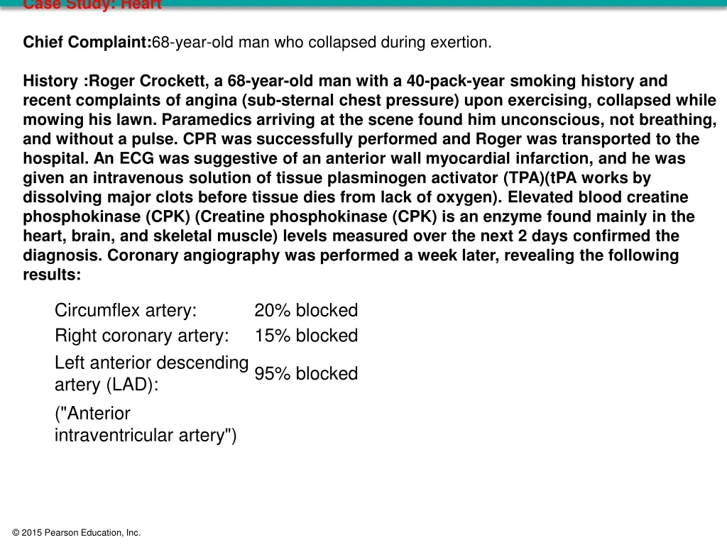
Case Study: Heart Chief Complaint:68-year-old man who collapsed during exertion. • History :Roger Crockett, a 68-year-old man with a 40-pack-year smoking history and recent complaints of angina (sub-sternal chest pressure) upon exercising, collapsed while mowing his lawn. Paramedics arriving at the scene found him unconscious, not breathing, and without a pulse. CPR was successfully performed and Roger was transported to the hospital. An ECG was suggestive of an anterior wall myocardial infarction, and he was given an intravenous solution of tissue plasminogen activator (TPA)(tPA works by dissolving major clots before tissue dies from lack of oxygen). Elevated blood creatine phosphokinase (CPK) (Creatine phosphokinase (CPK) is an enzyme found mainly in the heart, brain, and skeletal muscle) levels measured over the next 2 days confirmed the diagnosis. Coronary angiography was performed a week later, revealing the following results:
An Introduction to the Cardiovascular System • The PulmonaryCircuit • Carries blood to and from gas exchange surfaces of lungs • The SystemicCircuit • Carries blood to and from the body • Blood alternates between pulmonary circuit and systemic circuit
An Introduction to the Cardiovascular System • Three Types of Blood Vessels • Arteries • Carry blood away from heart • Veins • Carry blood to heart • Capillaries • Networks between arteries and veins
An Introduction to the Cardiovascular System • Capillaries • Also called exchangevessels • Exchange materials between blood and tissues • Materials include dissolved gases, nutrients, waste products
Pulmonary Circuit Systemic Circuit Systemic arteries Pulmonary arteries Systemic veins Pulmonary veins Figure 20-1 An Overview of the Cardiovascular System. Capillariesin head,neck, upper limbs Capillariesin lungs Rightatrium Leftatrium Rightventricle Leftventricle Capillariesin trunkand lower limbs
An Introduction to the Cardiovascular System • Four Chambers of the Heart • Rightatrium • Collects blood from systemic circuit • Rightventricle • Pumps blood to pulmonary circuit • Leftatrium • Collects blood from pulmonary circuit • Leftventricle • Pumps blood to systemic circuit
20-1 Anatomy of the Heart • The Heart • Great veins and arteries at the base • Pointed tip is apex • Surrounded by pericardial sac • Sits between two pleural cavities in the mediastinum
Trachea Thyroidgland First rib (cut) Figure 20-2a The Location of the Heart in the Thoracic Cavity. Base ofheart Left lung Right lung Parietalpericardium(cut) Apex ofheart Diaphragm a An anterior view of the chest, showing theposition of the heart and major blood vesselsrelative to the ribs, lungs, and diaphragm.
20-1 Anatomy of the Heart • The Pericardium • Double lining of the pericardial cavity • Visceralpericardium • Inner layer of pericardium • Parietalpericardium • Outer layer • Forms inner layer of pericardialsac
20-1 Anatomy of the Heart • The Pericardium • Pericardial cavity • Is between parietal and visceral layers • Contains pericardialfluid • Pericardial sac • Fibrous tissue • Surrounds and stabilizes heart
Base ofheart Cut edge ofparietal pericardium Figure 20-2c The Location of the Heart in the Thoracic Cavity. Wrist (correspondsto base of heart) Fibrous tissue ofpericardial sac Inner wall (corresponds to epicardium) Parietal pericardium Air space (corresponds to pericardial cavity) Areolar tissue Mesothelium Outer wall (correspondsto parietal pericardium) Cut edge of epicardium Fibrousattachment todiaphragm Balloon Apex of heart c The relationship between the heart and the pericardial cavity; compare with the fist-and-balloon example.
Base of heart 1 1 Ribs 2 2 Figure 20-3a The Position and Superficial Anatomy of the Heart. 3 3 4 4 5 5 6 6 7 7 Apex of heart 8 8 9 a 9 10 10 Heart position relative to the rib cage.
Left subclavian artery Left commoncarotid artery Arch of aorta Ligamentumarteriosum Brachiocephalictrunk Descendingaorta Ascendingaorta Left pulmonaryartery Figure 20-3b The Position and Superficial Anatomy of the Heart. Superiorvena cava Pulmonarytrunk Auricleof rightatrium Rightatrium Auricle ofleft atrium Fat and vesselsin anteriorinterventricularsulcus Rightventricle Fat andvessels incoronarysulcus Leftventricle Major anatomical features on the anterior surface. b
Arch of aorta Left pulmonary artery Right pulmonary artery Left pulmonary veins Fat and vesselsin coronarysulcus Superior vena cava Leftatrium Coronarysinus Figure 20-3d The Position and Superficial Anatomy of the Heart. Rightpulmonaryveins(superiorand inferior) Rightatrium Leftventricle Inferiorvena cava Rightventricle Fat and vessels in posteriorinterventricular sulcus d Major landmarks on the posterior surface. Coronaryarteries (which supply the heart itself) are shown in red; coronary veins are shown in blue.
20-1 Anatomy of the Heart • The Heart Wall • Epicardium • Myocardium • Endocardium
20-1 Anatomy of the Heart • Epicardium (Outer Layer) • Visceral pericardium • Covers the heart
20-1 Anatomy of the Heart • Myocardium (Middle Layer) • Muscular wall of the heart • Concentric layers of cardiac muscle tissue • Atrial myocardium wraps around great vessels • Two divisions of ventricular myocardium • Endocardium (Inner Layer) • Simple squamous epithelium
Pericardialcavity Parietalpericardium Myocardium(cardiac muscle tissue) Dense fibrous layer Cardiac muscle cells Areolar tissue Connective tissues Mesothelium Figure 20-4a The Heart Wall. Artery Vein Endocardium Epicardium(visceralpericardium) Endothelium Areolar tissue Mesothelium Areolar tissue Heart wall a A diagrammatic section through the heartwall, showing the relative positions of theepicardium, myocardium, and endocardium.The proportions are not to scale; thethickness of the myocardial wall has beengreatly reduced.
Atrialmusculature Figure 20-4b The Heart Wall. Ventricularmusculature Cardiac muscle tissueforms concentric layers thatwrap around the atria or spiralwithin the walls of the ventricles. b
20-1 Anatomy of the Heart • Cardiac Muscle Tissue • Intercalateddiscs • Interconnect cardiacmusclecells • Secured by desmosomes • Linked by gap junctions • Convey force of contraction • Propagate action potentials
Cardiac muscle cell Mitochondria Intercalateddisc (sectioned) Figure 20-5a Cardiac Muscle Cells. Nucleus Cardiac musclecell (sectioned) Bundles ofmyofibrils Intercalateddiscs Cardiac muscle cells a
Figure 20-5c Cardiac Muscle Cells. Intercalateddiscs LM x 575 Cardiac muscle tissue Cardiac muscle tissue c
20-1 Anatomy of the Heart • Characteristics of Cardiac Muscle Cells • Small size • Single, central nucleus • Branching interconnections between cells • Intercalated discs
Left common carotid artery Left subclavian artery Brachiocephalictrunk Ligamentumarteriosum Pulmonary trunk Superiorvena cava Aortic arch Pulmonary valve Rightpulmonaryarteries Left pulmonaryarteries Ascending aorta Figure 20-6a The Sectional Anatomy of the Heart. Left pulmonaryveins Fossa ovalis Leftatrium Opening ofcoronary sinus Interatrial septum Aortic valve Right atrium Cusp of left AV (mitral) valve Pectinate muscles Conusarteriosus Left ventricle Cusp of right AV(tricuspid) valve Chordae tendineae Interventricular septum Papillary muscles Trabeculaecarneae Right ventricle Inferior vena cava Moderator band Descending aorta A diagrammatic frontal section through the heart, showingmajor landmarks and the path of blood flow (marked byarrows) through the atria, ventricles, and associated vessels. a
Figure 20-6b The Sectional Anatomy of the Heart. Chordae tendineae Papillary muscles The papillary muscles and chordaetendineae support the right AV (tricuspid)valve. The photograph was taken frominside the right ventricle, looking towarda light shining from the right atrium. b
20-1 Anatomy of the Heart • The Heart Valves • Two pairs of one-way valves prevent backflow during contraction • Atrioventricular (AV) valves • Between atria and ventricles • Blood pressure closes valve cusps during ventricular contraction • Papillary muscles tense chordae tendineae to prevent valves from swinging into atria
20-1 Anatomy of the Heart • The Heart Valves • Semilunar valves • Pulmonary and aortic tricuspid valves • Prevent backflow from pulmonary trunk and aorta into ventricles • Have no muscular support • Three cusps support like tripod
20-1 Anatomy of the Heart • AorticSinuses • At base of ascending aorta • Sacs that prevent valve cusps from sticking to aorta • Origin of right and left coronary arteries
Transverse Sections, Superior View,Atria and Vessels Removed POSTERIOR Cardiacskeleton Left AV (bicuspid)valve (open) RIGHTVENTRICLE LEFTVENTRICLE Figure 20-8a Valves of the Heart (Part 1 of 2). Relaxed ventricles Right AV(tricuspid)valve (open) Aortic valve(closed) Pulmonaryvalve (closed) ANTERIOR a When the ventricles are relaxed, the AV valves are open and the semilunar valves are closed. The chordae tendineae are loose, and the papillary muscles are relaxed. Aortic valve closed
Frontal Sections through Left Atrium and Ventricle Pulmonaryveins LEFTATRIUM Figure 20-8a Valves of the Heart (Part 2 of 2). Left AV (bicuspid)valve (open) Chordaetendineae (loose) Relaxed ventricles Aortic valve(closed) Papillary muscles(relaxed) LEFT VENTRICLE(relaxed and fillingwith blood) a When the ventricles are relaxed, the AV valves are open and the semilunar valves are closed. The chordae tendineae are loose, and the papillary muscles are relaxed.
Left AV(bicuspid) valve(closed) Right AV(tricuspid) valve(closed) Cardiacskeleton LEFTVENTRICLE RIGHTVENTRICLE Figure 20-8b Valves of the Heart (Part 1 of 2). Contracting ventricles Aortic valve(open) Pulmonaryvalve (open) When the ventricles are contracting, the AV valves are closed and the semilunar valves are open. In the frontal section notice the attachment of the left AV valve to the chordae tendineae and papillary muscles. b Aortic valve open
LEFTATRIUM Aorta Aortic sinus Left AV (bicuspid)valve (closed) Aortic valve(open) Figure 20-8b Valves of the Heart (Part 2 of 2). Chordae tendineae(tense) Contracting ventricles Papillary muscles(contracted) Left ventricle(contracted) b When the ventricles are contracting, the AV valves are closed and the semilunar valves are open. In the frontal section notice the attachment of the left AV valve to the chordae tendineae and papillary muscles.
20-1 Anatomy of the Heart • The Blood Supply to the Heart • = Coronarycirculation • Supplies blood to muscle tissue of heart • Coronary arteries and cardiac veins
Aorticarch Left coronary artery Pulmonarytrunk Ascendingaorta Figure 20-9a The Coronary Circulation. Circumflexartery Rightcoronaryartery Anteriorinterventricularartery Atrialarteries Greatcardiacvein Anteriorcardiacveins Smallcardiac vein Marginal artery Coronary vessels supplying and draining the anteriorsurface of the heart. a
Coronary sinus Circumflex artery Great cardiac vein Marginal artery Posteriorinterventricularartery Figure 20-9b The Coronary Circulation. Posteriorcardiacvein Smallcardiacvein Leftventricle Rightcoronaryartery Marginal artery Middle cardiac vein b Coronary vessels supplying and drainingthe posterior surface of the heart.
Narrowing of Artery Normal Artery Figure 20-10 Heart Disease and Heart Attacks (Part 2 of 4). Tunicaexterna Lipid depositof plaque Tunicamedia Cross section Cross section
20-1 Anatomy of the Heart • Heart Disease – Coronary Artery Disease • Coronaryarterydisease (CAD) • Areas of partial or complete blockage of coronary circulation • Cardiac muscle cells need a constant supply of oxygen and nutrients • Reduction in blood flow to heart muscle produces a corresponding reduction in cardiac performance • Reduced circulatory supply, coronaryischemia, results from partial or complete blockage of coronary arteries
20-1 Anatomy of the Heart • Heart Disease – Coronary Artery Disease • Usual cause is formation of a fatty deposit, or atheroscleroticplaque, in the wall of a coronary vessel • The plaque, or an associated thrombus (clot), then narrows the passageway and reduces blood flow • Spasms in smooth muscles of vessel wall can further decrease or stop blood flow • One of the first symptoms of CAD is commonly anginapectoris
20-1 Anatomy of the Heart • Heart Disease – Coronary Artery Disease • Myocardialinfarction (MI), or heartattack • Part of the coronary circulation becomes blocked, and cardiac muscle cells die from lack of oxygen • The death of affected tissue creates a nonfunctional area known as an infarct • Heart attacks most commonly result from severe coronary artery disease (CAD)
20-1 Anatomy of the Heart • Heart Disease – Coronary Artery Disease • Myocardialinfarction (MI), or heartattack • A crisis often develops as a result of thrombus formation at a plaque (the most common cause of an MI), a condition called coronarythrombosis • A vessel already narrowed by plaque formation may also become blocked by a sudden spasm in the smooth muscles of the vascular wall • Individuals having an MI experience intense pain, similar to that felt in angina, but persisting even at rest
20-1 Anatomy of the Heart • Heart Disease – Coronary Artery Disease • Myocardialinfarction (MI), or heartattack • Pain does not always accompany a heart attack; therefore, the condition may go undiagnosed and may not be treated before a fatal MI occurs • A myocardial infarction can usually be diagnosed with an ECG and blood studies • Damaged myocardial cells release enzymes into the circulation, and these elevated enzymes can be measured in diagnostic blood tests • The enzymes include: • CardiactroponinT, • Cardiac troponin I, • A special form of creatinine phosphokinase, CK-MB
20-1 Anatomy of the Heart • Heart Disease – Coronary Artery Disease • Treatment of CAD and myocardial infarction • About 25 percent of MI patients die before obtaining medical assistance • 65 percent of MI deaths among those under age 50 occur within an hour after the initial infarction
20-1 Anatomy of the Heart • Heart Disease – Coronary Artery Disease • Treatment of CAD and myocardial infarction • Risk factor modification • Stop smoking • High blood pressure treatment • Dietary modification to lower cholesterol and promote weight loss • Stress reduction • Increased physical activity (where appropriate)
20-1 Anatomy of the Heart • Heart Disease – Coronary Artery Disease • Treatment of CAD and myocardial infarction • Drug treatment • Drugs that reduce coagulation and therefore the risk of thrombosis, such as aspirin and coumadin • Drugs that block sympathetic stimulation (propranolol or metoprolol) • Drugs that cause vasodilation, such as nitroglycerin • Drugs that block calcium movement into the cardiac and vascular smooth muscle cells (calcium channel blockers) • In a myocardial infarction, drugs to relieve pain, fibrinolytic agents to help dissolve clots, and oxygen
20-1 Anatomy of the Heart • Heart Disease – Coronary Artery Disease • Treatment of CAD and myocardial infarction • Noninvasive surgery • Atherectomy • Blockage by a single, soft plaque may be reduced with the aid of a long, slender catheter inserted into a coronary artery to the plaque
20-1 Anatomy of the Heart • Heart Disease – Coronary Artery Disease • Treatment of CAD and myocardial infarction • Noninvasive surgery • Balloonangioplasty • The tip of the catheter contains an inflatable balloon • Once in position, the balloon is inflated, pressing the plaque against the vessel walls • Because plaques commonly redevelop after angioplasty, a fine tubular wire mesh called a stent may be inserted into the vessel, holding it open
20-1 Anatomy of the Heart • Heart Disease – Coronary Artery Disease • Treatment of CAD and myocardial infarction • Coronary artery bypass graft (CABG) • In a coronary artery bypass graft, a small section is removed from either a small artery or a peripheral vein and is used to create a detour around the obstructed portion of a coronary artery • As many as four coronary arteries can be rerouted this way during a single operation • The procedures are named according to the number of vessels repaired, so we speak of single, double, triple, or quadruple coronary bypasses
Figure 20-10 Heart Disease and Heart Attacks (Part 1 of 4). Advanced Coronary Artery Disease A color-enhanced DSA scan showingadvanced coronary artery disease. Bloodflow to the ventricular myocardium isseverely restricted. Normal Heart A color-enhanced digitalsubtraction angiography (DSA)scan of a normal heart.
Figure 20-10 Heart Disease and Heart Attacks (Part 3 of 4). OccludedCoronaryArtery DamagedHeartMuscle
20-2 The Conducting System • Heartbeat • A single contraction of the heart • The entire heart contracts in series • First the atria • Then the ventricles


