

SlideServe
Menu
Préférences
© SlideServe · v2026

WELCOME

stefan avatar
24 diapositives · 966 Views · Sep 01, 2014

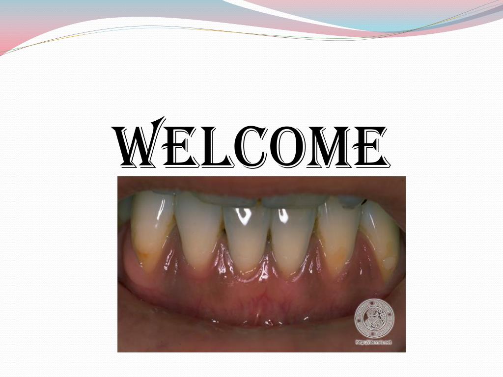
WELCOME. PIGMENTED LESIONS OF ORAL MUCOSA. Oral and Perioral pigmentation may be physiologic (or) pathologic in origin. Assume variety of discolorations, including brown,blue, grey & black.
WELCOME - slide 1
01 / 24WELCOME
1 / 17

Télécharger la présentation

WELCOME
Une image / un lien ci-dessous est fourni (tel quel) pour télécharger la présentation

Politique de téléchargement : le contenu du site vous est fourni TEL QUEL pour votre information et votre usage personnel ; il ne peut être vendu, concédé sous licence ni partagé sur d’autres sites sans l’accord de son auteur.Le contenu vous est fourni TEL QUEL, pour votre information et votre usage personnel uniquement. Téléchargez la présentation en cliquant sur ce lien. Lors du téléchargement, si vous ne parvenez pas à télécharger une présentation, l’éditeur a peut-être supprimé le fichier de son serveur.Pendant le téléchargement, si vous ne parvenez pas à obtenir une présentation, le fichier a peut-être été supprimé par l’éditeur.

F I N

Plus de contenu associé

Transcription de la présentation
  1. WELCOME

  2. PIGMENTED LESIONS OF ORAL MUCOSA • Oral and Perioral pigmentation may be physiologic (or) pathologic in origin. • Assume variety of discolorations, including brown,blue, grey & black. • These color changes often occur due to deposition,production (or) increased accumulation of various endogenous (or) exogenous pigmented substances.

  3. CLASSIFICATION • ENDOGENOUS PIGMENTATION • FOCAL MELANOCYTIC PIGMENTATION • Freckle / Ephelis • Oral / labial melanotic macule • Oral melanoacanthoma • Melanocytic nevus • Malignant melanoma • MULTIFOCAL / DIFFUSE PIGMENTATION • Physiologic pigmentation • Drug induced melanosis • Smoker melanosis • Post inflammatory hyper pigmentation • Melasma (chlosma)

  4. MELANOSIS ASSOCIATED WITH SYSTEMIC (OR)GENETIC DISEASE • Hypo adrenocorticism (addison’s disease) • cushing’s syndrome • Hyperthyrodism (graves disease) • Primary biliary cirrhosis • Vitamin b12 deficiency • Peutzjeghers syndrome • Café au lait pigmentation • HIV / AIDS associated melanosis

  5. IDIOPATHIC PIGMENTATION Laugier – hunziker pigmentation • DEPIGMENTATION Vitiligo • HAEMOGLOBIN & IRON ASSOCIATEDPIGMENTATION • Ecchymosis • Purpura / Petechiae • Hemochromatosis

  6. EXOGENOUS PIGMENTATION

  7. Focal melanocytic pigmentation • Freckle / ephelis : The cutaneous freckle is a commonly occuring, asymptomatic , small (1 – 3mm), well circumsribed , tan or brown colored macule that is often seen on sun exposed regions of facial & perioral skin. Polymorphisms in mc1r gene is strongly associated with development of childhood freckles.

  8. Oral / labial melanotic macule • Etiology is trauma • More commopn in females usually in lower lip &gingiva. • May develop at any age but generally tend to present in adulthood. • Melanotic macules tend to be small (<1 cm), well circumscribed , oval or irregular in outline & often uniformly pigmented.

  9. Oral melanoacanthoma • Etiology is acute trauma or a history of chronic irritation • Rapidly enlarging , ill defined , darkly pigmented macular or plague like lesions. • Buccal mucosa most common site of occurrence • Dermatosis papulosa nigra is relatively common facial condition in older black females & represents multiple pigmented seborrheic keratoses • Treatment is source of irritation should be removed

  10. Melanocytic nevus • Effect of sun exposure reconized in development of cutaneous nevi. • Recent study shows 90% of dermal melanocytic nevi exhibit somatic activating mutations in BRAF oncogene. • Lesions are usually asymtomatic & often present as a small (<1cm) , solitary , brown or blue , well circumscribed nodule or macule. • Oral nevi present in hard palate is the most common site , followed by buccal & labial mucosa & gingiva.

  11. Treatment of melanotic nevi is complete but conservative surgical excision is the treatment of choice for oral lesions. • Rare recurrence • Laser & intense pulse light therapies have been used successfully

  12. Malignant melanoma • Etiology is episodes of acute sun exposure , especially at young age , immunosuppresion • Exhibit mutation in the BRAF , HRAS & NRAS proto oncogenes. • Palate represents most common site & next comes the maxillary gingiva. • They are macular , plague like or mass forming , well circumscribed or irregular & exhibit focal or diffuse areas of brown , blue or black pigmentation.

  13. Treatment is ablative surgery with wide margins . • Adjuvant radiotherapy is necessary. • Recent development is antitumor vaccine adjuvant interferon alpha – 2B therapy is used to treat primary cutaneous melanomas >4mm in thickness.

  14. Multifocal / diffuse pigmentation • Physiologic pigmentation is more common • Mostly occur in gingiva • Treatment is gingivectomy & laser therapy have been used to remove pigmenteds oral mucosa. • Effects of treatment is temporary & may eventually recur.

  15. Drug induced melanosis Chief drugs include • Minocycline – tetracycline derivative • Antimalarials include chloroquine , hydroxy chloroquine , quinacrine . • Phenothiazines such as chlorpromaqzine • Oral contraceptives • Cytotoxic medications such as cyclophosphamide & busulfan

  16. Clinically the pigment can be diffuse yet localised to one mucosal surface , often hard palate • Lesions are flat & without any evidence of nodularity or swelling. • Diagnosis & treatment is discolaration tends to fade within a few months after the drug is discontinued.