
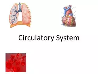
Circulatory System
The circulatory system plays a crucial role in maintaining bodily functions, consisting of blood, the heart, and blood vessels. Blood, a connective tissue, comprises plasma (55%) and formed elements (45%), including red blood cells, white blood cells, and platelets. Each component has vital functions: transportation of nutrients and waste, regulation of body temperature and pH, and protection against infections. Disorders like anemia, leukemia, and hemophilia can affect blood function, while blood types (ABO and Rh) influence compatibility for transfusions.

Circulatory System
E N D
Presentation Transcript
Circulatory System • Consists of the blood, heart, and blood vessels
Blood • Type of Connective tissue • 4-6 liters in the body • Has a liquid and solid portion.
Liquid portion: Plasma (55%) • (mostly water) • Solid portion: platelets, Red blood cells, and White blood cells (45%) • Buffy coat (wbc and platelets) less than 1% • The bottom is red blood cells
The “formed elements” of blood • Erythrocytes (red blood cells) • Leukocytes (white blood cells) • Thrombocytes (platelets)
Functions of Blood • Transportation- waste nutrients, and hormones • Regulation- body temp, pH • Protection- blood clots to prevent blood loss and fights infections
Plasma • Extra cellular matrix in blood • Mostly water (>90%) • Proteins (7%) produced by the liver • Albumin- helps with water balance in blood stream • Globulin- transports HDL and LDL proteins • Fibrinogen – clotting proteins • Other solutes such as oxygen, carbon dioxide, urea
Structure of a Erythrocyte • Small (7micrometers by 2 micrometers) • Biconcave • Flexible • No nucleus or organelles • “ghost of a cell” • Packed with hemoglobin • *2 ½ trillion in one person*
Hemoglobin carries oxygen • Oxygen binds to the iron containing heme group • Some carbon dioxide will bind as well
Oxygen loading and unloading • Oxygen is loaded onto the heme in the lungs forming oxyhemoglobin (bright red) • Oxygen is unloaded in the tissues forming deoxyhemoglobin (darkish red to purple)
Lifespan of Erythrocytes • Made in the red marrow of the long bone • Live for about 120 days • Dead RBCs go to the spleen were they are decomposed
Anemia • Reduction in oxygen carrying capacity • Measured as hematocrit- percentage of blood volume made up by RBCs (should be 45%) • Symptoms: Tired, bruising easily • Types of anemia • Iron deficiency • Hemorrhagic (bleeding) • Hemolytic (breaking RBC) • Sickle Cell (genetic disorder) • CO poisoning (takes place of O)
Sickle Cell Anemia • Many RBCs have a Sickle like shape • Genetic Recessive disorder • You can be a carrier or actually have sickle cell anemia • Thought to be a defense against malaria
Polycythemia • Excess of RBCs in blood • Increases blood viscosity (difficult to circulate blood)
White Blood Cells (WBC) • “Leukocytes” • Fewer in number (5,000-10,000 in the body) • True cells with nuclei and organelles • Guard/Fight Infections • Larger than RBCs • Found in the “Buffy Coat”
2 different classes of WBCs • Granular Leukocytes (Granulocytes) • Agranular Leukocytes (Agranulocytes)
Granular leukocytes • Neutrophils- fight bacterial infection • Eosinophils- fight parasitic infection • Basophils- release histamines, allergic responses
Agranular Leukocytes • Lymphocytes- • T lymphocytes attack virus infected cells and tumors • B lymphocytes produce antibodies • Monocytes- largest WBC, phagocytize, high with chronic infections
Leukemia • Uncontrolled reproduction of WBCs • Named for abnormal cell type • Myelocytic leukemia – uncontrolled granular line • Lymphocytic leukemia- uncontrolled agranular line
Mononucleosis • Caused by ebstein-barr virus • Excessive numbers of agranulocytes and basophils
Platelets • “Thrombocytes” • Important in Clotting
Hemostasis: Stopping Blood loss • Tear a blood vessel due to a cut or scrape • Positive feedback cycle • Platelets form a temporary plug • Coagulation- formation of clot with fibrin strands
Hemophilia • Blood is not able to clot properly because its missing a clotting factor • Bleed for a long time • located on the X chromosome • females can carry and pass to sons
Blood Typing • Erythrocytes carry antigens on their surface • Antigens make the immune system respond • Antigens are proteins • Plasma contains antibodies that will attack FOREIGN antigens
The ABO Blood System • 4 Types: A, B, AB, and O • Each type has specific antigens on the RBC surface • specific antibodies in the plasma
The ABO Blood System • Type A blood, has A antigens on the red blood cell’s surface. • Type B blood will have B antigens. • Type AB blood will have A and B antigens • Type O blood IS THE EXCEPTION, it has NO antigens on its surface
Antibodies • Located in the plasma • protect body against foreign substances INCLUDING foreign blood types
The ABO Blood System • Blood type AB is the most rare. • Blood type O is the most frequent blood type.
The ABO Blood System • Type O- is considered to be a “universal donor” • Type AB+ is considered to be a “universal recipient”
Rh Factor • Another way to classify blood • “Rhesus Factor” • Rh+ is positive for the Rh protein on RBC surface • Rh- negative for the Rh protein on RBC surface
Rh Factor • people with Rh+ blood will not have Rhantibodies • People will Rh- blood will have Rhantibodies in plasma • react by coagulating when exposed to Rh+ blood.
Hemolytic disease of the newborn • Seen with children who are Rh+ and their mother is Rh- • Blood is transferred through the placenta, mother develops strong antibodies against + • 1st child will be fine, next child is at risk from mom’s antibodies attacking baby • Rhogam is given to the mother to protect baby
Transfusions • Transfusions- blood from one person is taken OUT of their body and PUT into another person’s Body.
Transfusion Reaction • Occurs when antibodies attack foreign RBCs that have entered the body • Coagulation • Heart attack, stroke, renal failure may result
The Heart • Provides the force to propel blood through the system • Located in thoracic cavity, medial to the lungs, anterior to the vertebrae, and posterior to the sternum
Two Circuits • Heart is involved in the two circuits of the circulatory system. • Pulmonary • Heart, lungs, Heart • Systemic • Heart, body, heart
Stats of the heart • Size: clenched fist • Shape: Cone shaped • Pointed apex, points toward left and rests on diaphragm
Surroundings • Surrounded by the pericardial cavity • Parietal pericardium: Lines the PERICARIDAL SAC • Visceral pericardium “epicardium” covers the OUTER SURFACE of the heart
Layers of the Heart From OUT to IN • Epicardium: covers outer surface • Myocardium: muscle (walls) • Endocardium: lining of the inside of the heart
Chambers of the Heart • 2 Atria and 2 Ventricles • Atria- superior; RECEIVE blood, thin walls • Ventricles- inferior; PUMP blood, thick walls
Atria • Small and ear like in shape. Located superiorly. • There is a right and a left • The right receives deoxygenated blood from the body • The left receive oxygenated blood from the lungs.
The Heart- Ventricles • take up the majority of the heart. • Left is largest and most muscular • right receives deoxygenated blood from the right atrium and pumps out to lungs • left receives oxygenated blood from the left atria and pumps out to body
Valves • All 4 Valves prevent back flow of blood • 2 Atrioventricular (AV) valves prevent blood from coming back out of the ventricles and into the atria • 2 Semilunar values prevent backflow from the great vessels into the ventricles
AV valves • Bicuspid- between Left atria and left ventricle • Tricuspid- between right atria and right ventricle • Attached to chordaetendinae “Heart stings” which are attached to papillary muscles
Semilunar Valves • Aortic valve and pulmonary valve • Aortic semilunar valve separates the left ventricle and aorta • Pulmonary semilunar valve separates the right ventricle and the pulmonary artery


