Download
1 / 38
380 likes | 416 Vues
Dive into the intricate world of the nervous system - the primary communication and control center of the body. Learn about neurons, neuroglia, and the divisions of the nervous system. Explore the brain's functions and structure to grasp its essential role in maintaining homeostasis.

E N D
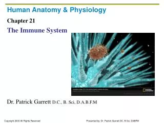
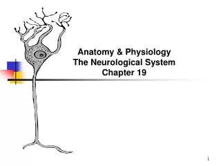
Anatomy & Physiology The Neurological System Chapter 19
The nervous system takes impressions and stimuli from the outside world and selectively stores information for the future. The Nervous system also coordinates the internal body systems and allows the body to constantly readjust to constantly changing internal and external environments. Nerves are like wires that carry ingoing and outgoing messages. • Neurology is the study of the nervous system. A neurologist is someone who studies neurology.
Structure and Function • The main function of the nervous system is communication and control. • Communication • Monitors impressions and information from external stimuli • Monitors information from internal stimuli • Responds to danger, pain and other situations • Responds to internal and external changes • Helps maintain homeostasis • Responds to conscious decisions and thoughts • Coordinates the process of new learning • Control • Directs all body activities • maintains blood pressure, respiration and other vital functions • Regulates body systems • coordinates reflexes • Controls instinctual behavior • Controls conscious movement and activities • Stores unconscious thoughts
The Cells of the Nervous System • Two Types: • The Neuron- the basic structural and functional cell of the nervous system. • The Neuroglia - five times more numerous than neurons. They do not transmit impulses, but support and connect nervous tissue.
The Neurons • Has many functions and vary in size and in length. • Three basic parts to a Neuron • cell body • axon • dendrites • Each Neuron has one cell body with a nucleus. Neurons cannot divide and multiply by mitosis like other cells in the body. Once the body is destroyed it is gone forever. • The axon is an extension that carries impulses away from the neuron cell body. Some have a covering called myelin and others do not. • Myelin sheath is a fatty covering. Those axons that have myelin are called myelinated axons. Myelinated axons conduct impulses more rapidly than unmyelinated axons. These sheaths help to insulate the nerve cell and conduct impulses easily and rapidly. • Dendrites are the short, highly branched parts of the cell body. They carry impulses from the axon and send and receive impulses across the synapse. • A synapse is the junction or space between the axon of one neuron and the dendrites of another.
A nerve can only transmit impulses in only one direction because of the location of neurotransmitters. These are chemicals that the axon releases to allow nerve impulses to cross the synapse and reach the next nerves dendrites. The dendrites release opposing chemicals to slow down impulses. • Neurons can be classified as follows: • Sensory ( afferent) neurons- receive and send messages to the central nervous system from all parts of the body. • Motor ( efferent) – neurons receive and transmit messages from the central nervous system to all parts of the body. • Interneurons (connectory/ association neurons/ or integrators) can be thought of as a link between the two other types of neurons. They are interconnecting neurons. • Sensory neurons make up sensory nerves. • Motor neurons make up motor nerves- which cause muscle activity and gland secretion. • Put together, sensory and motor neurons make up Mixed nerves.
Neuroglia • Also known as glial cells are more numerous than neurons. • They can multiply to fill spaces previously inhabited by neurons. • There are four types of neuroglia in the central nervous system: • astrocytes • oligodendrocytes • microglia • ependymal cells • There are two type of neuroglia in the peripheral nervous system: • neurolemmocytes • Schwann Cells. • These two types of cells help form the blood-brain barrier- the cerebrospinal fluid- and the myelin sheath. • They also help to obtain nutrients for the neurons and act as support and protection for the nervous system overall.
Divisions of the Nervous System • Central Nervous System- consists of the brain and the spinal cord and other accessory structures. • Peripheral Nervous System- consists of the cranial nerves, spinal nerves, and the autonomic nervous system.
The Central Nervous System. THE BRAIN • The Brain is composed of approximately 100 billion neurons and synapses. It weighs about 3 lbs, and that is about 2% of the body weight. The brain has an extensive vasculature. It works faster than the rest of the body ( higher metabolic rate). The brain must have a constant flow of oxygen and glucose or it will begin to shut down immediately. The brain has many parts that are integrated.
Cerebrum- is the largest part of the brain (80%). It is divided into two layers and two halves (hemispheres). Each portion of the cerebrum has its own specialized function. • Cerebral Cortex- points to the unique human abilities of learning, intelligent reasoning, and judgment. This is the outside layer of the brain. • It is made up of soft, gray matter, mostly nerve cell bodies. There are wrinkles and folds called convolutions or gyri. The crevices between the folds are called fissures or sulci. This gives the cortex a larger surface area to hold more neurons. The cerebral cortex is further subdivided into four lobes • Frontal lobe- Controls areas for written and motor speech. Helps with conception, judgment, speech, and communication. It is involved with motor function that directs body movements. • Parietal lobe- is the sensory area because this is where touch, temperature, pain sensation emanate. Spatial ability also located in this area. • Temporal lobe- controls the sensation of hearing, auditory interpretation, and smell. • Occipital lobe- helps with visual transmission and interpretation. • White Matter- is in the interior of the brain. It is under the cerebral cortex. It is white because of the mass amounts of white myelinated axons that connect the lobes of the cerebrum together. • Cerebral hemispheres- the right hemisphere of the brain controls the muscles of and receives impulses from the left side of the body and vice versa. This phenomena is called decussation (crossing) of the nerve tracts within the brain’s medulla. The two hemispheres process information differently. • The right side is associated with spatial perception, pictures, art and musical ability. • the left side is associated with analytic and verbal skills and walking. • The Corpus Callosum is a band of 200 million neurons located deep within the brain. It connects the right and left side of the brain. It allows both sides to share information and to integrate that information.
Thalamus- is located in the diencephalon portion of the brain, between the hemispheres and the brain stem. It lies just superior to the hypothalamus. It is the relay system between the cutaneous receptors and the cerebral cortex for all sensory impulses, except smell. The thalamus integrates the sensations so a person can feel a whole experience rather than just individual impulses. Ie: Touching a snowball
Hypothalmus- is vital to human functioning. It is located below the thalamus in the diencephalon portion of the brain. Most functions of the hypothalamus are related somewhat to visceral activities. It helps increase or decrease body functions, regulates the release of hormones from the pituitary gland ( temperature control, water balance, sleep, appetite, emotions).
Limbic System- is located between the cerebrum and the inner brain. Responsible for maintaining a sense of awareness. • Hippocampus- functions for learning and long term memory • Reticular formation- governs senses coming into the cortex as well as sleep and wakefulness
Cerebellum- is the second largest portion of the brain. It’s functions are concerned with movement: muscle tone, coordination, and equilibrium. It helps coordinate the functions of voluntary muscles to allow for balance, walking, dancing, skating
Brain Stem- connects the cerebral hemispheres with the spinal cord. It includes the midbrain, the pons, and the medulla. • The midbrain is located at the very top of the brain stem and acts as a reflex center. Visual and auditory reflexes are integrated here. When you turn your head to locate a sound, you are using the midbrain. The ‘righting reflex’ or the ability to hold your head upright and maintain balance is also found here. • The pons- contains nerve tracts that carry messages between the cerebrum and the medulla. The pons has respiratory centers that work with the medulla to produce normal breathing patterns. • The medulla is just below the pons and rests on the floor of the skull. It is continuous, but not part of the spinal cord. The cardiac center, vasomotor center, the repiratory center are located here. The nerve tracts from the cerebrum cross over here as they enter the spinal cord. Swallowing, coughing, sneezing.
The Spinal Cord • Is a long mass of nerve cells and fibers extending through a central canal from the medulla to the first or second lumbar vertebra. It has two main functions: • To conduct impulses to and from the brain (Reflex arcs). • To act as a reflex center. Some nerves are sensory, in that they carry messages to the brain and some are motor, in that they carry messages away from the brain.
Accessory Structures • There are three accessory structures of the Central Nervous System. • meninges • cerebrospinal fluid • ventricles
Meninges • - The brain and the spinal cord are covered with a protective layer called the meninges. It is in 3 layers. • The dura mater- is the outer layer and is tough and fibrous and connects to the bones of the skull. • The arachnoid- is the middle layer. • The Pia mater- is the inner layer that lies closely over the spinal cord. It has many blood vessels that help bring oxygen and nutrients to the spinal cord. • The space between the pia mater and the arachnoid mater is called the subarachnoid space and it is here that we find cerebrospinal fluid. • *Head trauma can result in intracranial bleeds with blood accumulation under specific meninges: • Subdural (under the dura mater) • Subarachnoid (under the arachnoid)
b. Cerebrospinal Fluid- • is a lymph like fluid that forms a protective cusion around and within the CNS. CSF allows the brain to float within the cranial cavity. It removes cellular waste and lessens the impact of trauma. It is produced in specialized networks of the ventricles of the brain called the choriod plexuses. About 800 milliliters are produced daily, although only about 200 circulate at any given time. • Lumbar puncture- a physician withdraws fluid for analysis, introduction of medication and measuring CSF pressure. • Increased CSF pressure can be a sign of serious disorder such as brain tumor, bleeding, hydrocephalus or infection.
Ventricles • There are four ventricles or cavities deep within the brain. They are lined with ependymal cells and contain blood vessels from the pia mater, which make up the choroid plexuses. This is where CSF is made and fills the ventricles.
The Peripheral Nervous System • Is generally made up of two nerve groups. Cranial Nerves and Spinal Nerves. • Cranial Nerves- are nerves that begin in the brain • Spinal- are nerves that begin in the spine. The nerves of the Peripheral Nervous System are either sensory, motor, or mixed, depending on which direction the impulses are conducted. The autonomic nervous system is also classified as part of the PNS.
The Cranial Nerves • There are 12 pairs of cranial nerves that attach directly to the brain. Most carry impulses to and from the brain in various structures around the head. One pair acts on the organs of the thorax and the abdomen. • 12 nerves- ON OLD OLYMPUS, TOWERING TOP A FINN AND A GERMAN VIEW SOME HOPS. • Function of the 12 nerves- SOME SAY MARRY MONEY BUT MY BROTHER SAYS BAD BUSINESS MARRY MONEY. • S= SENSORY • B=BOTH • M=MOTOR • All the cranial nerves are important. However, the vagus nerve should be discussed. • The vagus nerve is responsible for many of the unconscious functions of the body. Pharynx, larynx, respiratory tract, heart, esophagus, and parts of the abdominal viscera are controlled by the vagus nerve.
The Spinal Nerves • There are 31 pairs of spinal nerves attached to the spinal cord. Each group of spinal nerves is named for its corresponding part of the spinal cord: • Cervical- 8 pairs • Thoracic- 12 pairs • Lumbar- 5 pairs • sacral-5 pairs • Coccygeal- 1 pair. • Each spinal nerve has a dorsal/posterior ( section that receives sensory information) and a ventral/anterior ( section that carries motor impulses to muscles and glands. • A group of spinal nerves is called a plexus • Phrenic nerve- controls the diaphragm • Brachial plexus- nerves to the upper arms • Lumbosacral plexus- where the sciatic nerve arises • Pudendal plexus- nerves from the perineum arise. • An injury to any of these plexus can lead to nerve damage. Pain medication may numb some of these plexus to reduce the sensation of pain in a particular area.
The Autonomic Nervous System • Is composed of portions of the CNS and PNS. It functions independently and without conscious effort. It innervates muscle that is not under conscious control like cardiac muscle, smooth (visceral) muscles and glands. The ANS contains visceral motor neurons that help transmit impulses to these three areas. It is further subdivided into the sympathetic and parasympathetic systems. • The Sympathetic Division • Is the part of the ANS that causes the body to respond in an emergency. Senses are greatly increased to respond to stimuli. • The Parasympathetic Division • Generally produces normal body functions of the body while it is at rest and brings the body back to normal after an emergency.
Physiology of the Nervous System Transmission of Nerve Impulses • Messages from one part of the body can take several different pathways. However, the body will tend to use the quickest method possible to complete an impulse. The body picks up habits by using the same nervous pathway repeatedly. Repeated motions become more or less automatic. • Action Potential- a neuron receives electrical and chemical impulses, which make it possible for the neuron to transfer a stimulus from one area of the body to another and to elicit a response. The electrical impulse is due to the positive and negative charged electrolytes. • A rapid exchange of sodium and potassium ions takes place when there is an impulse. The impulse moves across these ions in a millisecond along a nervous pathway. • At the synapse, neurotransmitters act chemically to transfer an impulse from the axon of one neuron to the dendrites of another. As chemicals are released, the polarity of the first cell is reversed to normal and the cell returns to its resting state.
EEG is a measurement of these brain waves or electrical activity of millions of brain cells.
Actions of the three types of Neurons. • Sensory Neurons are also called afferent neurons because they carry impulses to the brain or spinal cord from the periphery of the body by means of receptors. Receptors are end organs that initially receive stimuli from outside and within the body. • Exteroceptors- related to the external body • Proprioceptors- carry the sensation of position and balance. • Interoceptors- respond to changes in the internal organs. • When the receptors have picked up an impulse, fibers of the sensory neurons carry the sensation to the CNS.
Motor Neurons are also called efferent neurons because they carry impulses away from the CNS. Causes the body to respond to stimuli. • Interneurons- assist with thinking, learning, and memory and can be though of as a link between sensory and motor neurons.
Reflex • Is an automatic or involuntary response to a stimulus. Reflexes occur without conscious thought. They are controlled by the CNS and can be exaggerated or dulled according to the need of the body. Constriction of the pupils when exposed to light, increase in heart rate when the body senses a lowering in blood pressure. Knee jerk when the examiner taps the patellar tendon.
Effects of Aging on the System • Intelligence, memory, the capacity to learn, and personality do not normally change as a person ages. Nerve cells cannot reproduce themselves. Damage to brain cells can result in permanent loss in mental functioning. Strokes, Alzheimer’s disease, Parkinson’s disease effect brain function. Loss of equilibrium and changes in proprioception ( spatial awareness) can contribute to falls. Herpes Zoster is a painful nerve disorder in older adults.