Download
1 / 45
450 likes | 835 Vues
Introduction to Cells (Cytology Part I). Text: BJ Chap 3, AP Chap 6 Reading Assignments J Chap 3 pp 69– 94, AP pp167 - 192Text: BJ Chap 2, AP Chap 5 Homework Assignment Chap 3 Review Questions. BJ 3A – The structure of Cells (p 69). Hierarchy of Life

E N D
Introduction to Cells (Cytology Part I) • Text: BJ Chap 3, AP Chap 6 • Reading Assignments • J Chap 3 pp 69– 94, AP pp167 - 192Text: BJ Chap 2, AP Chap 5 • Homework Assignment • Chap 3 Review Questions
BJ 3A – The structure of Cells (p 69) • Hierarchy of Life • Atoms -> Molecules -> Cell Structures -> Organs -> Organ Systems
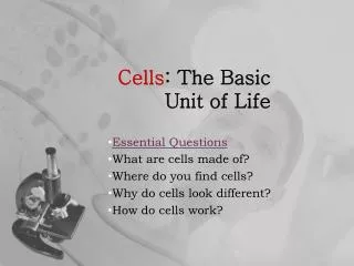
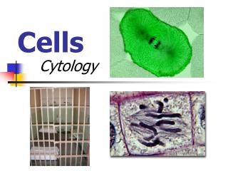
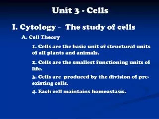
BJ 3 A- 1. (p 70) Cell Theory • Cytology:Cytology means "the study of cells". Cytology is that branch of life science, which deals with the study of cells in terms of structure, function and chemistry. Based on usage it can refer to: • Cell biology - the study of (normal) cellular anatomy, function and chemistry. • Cytopathology - the study of cellular disease and the use of cellular changes for the diagnosis of disease.
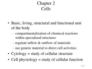
Cell as Basic Unit • Cell: The cell is the structural and functional unit of all known living organisms. It is the smallest unit of an organism that is classified as living, and is often called the building block of life. Some organisms, such as most bacteria, are unicellular (consist of a single cell). Other organisms, such as humans, are multicellular. (Humans have an estimated 100 trillion or 1014 cells; a typical cell size is 10 µm; a typical cell mass is 1 nanogram.) The largest known cell is an unfertilized ostrich egg cell.
Protoplasm: • Protoplasm is the living contents of a cell that are surrounded by a plasma membrane. • This term is not commonly used in modern cell biology. • Protoplasm is composed of a mixture of small molecules such as ions, amino acids, monosaccharides and water, and macromolecules such as nucleic acids, proteins, lipids and polysaccharides. • In eukaryotes the protoplasm surrounding the cell nucleus is known as the cytoplasm and that inside the nucleus as the nucleoplasm. In prokaryotes the material inside the plasma membrane is the bacterial cytoplasm, while in gram-negative bacteria the region outside the plasma membrane but inside the outer membrane is the periplasm. • Protoplasm is distinct from non-living cell components lumped under "ergastic substances" or inclusion bodies, although ergastic substances can occur in the protoplasm. In many plant cells most of the volume of the cell is not occupied by protoplasm, but by "tonoplast," a large water filled vacuole enclosed by a membrane. A protoplast is a plant or fungal cell that has had its cell wall removed.
Cell Theory: • Cell Theory: refers to the idea that cells are the basic unit of structure in every living thing. Development of this theory during the mid 1600s was made possible by advances in microscopy. This theory is one of the foundations of biology. The theory says that new cells are formed from other existing cells, and that the cell is a fundamental unit of structure, function and organization in all living organisms.
Cell as a Functional Unit (AP 167) • (Table 3A-1 p 71) • The cell performs all of the major functions of all living things. Examples: • Nutrition: Absorption - transport of dissolved substances (nutrients) into the cells • Digestion - Breakdown of nutrient with enzymes. • Internal Functions: Synthesis, Respiration, Movement, Irritability • Material Release: Excretion - removal of soluble wastes • Egestion - Elimination of non soluble wastes • Secretion - Synthesis and release of substance from a cell • Continuation of Existence: Homeostasis - keeping steady state in the cell • Reproduction • Cell as a Reproductive Unit • Cells reproduce by dividing.
BJ 3A-2 Levels of Cellular Organization (BJ 73 – 74) • Unicell organism: Most microorganisms are single-celled, or unicellular. Some are so small they are microscopic, but some unicellular protists are large enough to be visible to the average human. (See cell size table BJ page 72) • Multicell organisms: composed of more than one cell, typically many cells. • Colonial organisms: A collection of similar cells living together. Each cell except for reproductive cells can carry on all process of organism if separated by the colony. • Tissue: group of similar cells that work together to perform a specific function. • Organs: An organ is a fully differentiated structural and functional unit composed of one or more tissue types in an animal that is specialized for some particular function or functions. • Organ system: A group of organs that work together to perform a certain task.
See BJ Table 3A-2 page 77. • 3A-3 Cellular Anatomy • Three broad categories of cell anatomy: • Boundary the encloses the cell • Cytoplasm containing various kinds of structures and molecules • Nucleus that contains DNA and other materials
Two Types of Cells: • Eukaryotic: A is an organism whose cells contain complex structures enclosed within membranes. Most living organisms, including all animals, plants, fungi, and protists, are eukaryotes. The defining membrane-bound structure that differentiates eukaryotic cells from prokaryotic cells is the nucleus. The presence of a nucleus gives these organisms their name, which comes from the Greek ευ (eu, "good", "true") and κάρυον (karyon, "nut"). Many eukaryotic cells contain other membrane-bound organelles such as mitochondria, chloroplasts and Golgi bodies. • Cell division in eukaryotes is different from organisms without a nucleus (prokaryotes). It involves separating the duplicated chromosomes, through movements directed by microtubules. There are two types of division processes. In mitosis, one cell divides to produce two genetically identical cells. In meiosis, which is required in sexual reproduction, one diploid cell (having two instances of each chromosome, one from each parent) undergoes recombination of each pair of parental chromosomes, and then two stages of cell division, resulting in four haploid cells (gametes). Each gamete has just one complement of chromosomes, each a unique mix of the corresponding pair of parental chromosomes. • Eukaryotes appear to be monophyletic, and so make up one of the three domains of life. The two other domains, Bacteria and Archaea, are prokaryotes and have none of the above features. • Organelle: a cytoplasmic structure within the cell that performs special functions within the cells.
Prokaryotic: The prokaryotes (singular pronounced /proʊˈkæriət/) are a group of organisms that lack a cell nucleus (= karyon), or any other membrane-bound organelles. They differ from the eukaryotes, which have a cell nucleus. Most are unicellular, but a few prokaryotes such as myxobacteria have multicellular stages in their life cycles.[ It is also spelled "procaryote". • The prokaryotes are divided into two domains: the bacteria and the archaea. Archaea were recognized as a domain of life in 1990. These organisms were originally thought to live only in inhospitable conditions such as extremes of temperature, pH, and radiation but have since been found in all types of habitats.
Cell Boundaries (BJ 77) • Cell boundaries include the plasma membrane which is part of the cell, and the cell walls, capsules and sheaths which are on the outside of the cell itself. • Plasma Membrane: The cell membrane (also called the plasma membrane or plasmalemma) is the biological membrane separating the interior of a cell from the outside environment. It is a semipermeable lipid bilayer found in all cells. It contains a wide variety of biological molecules, primarily proteins and lipids, which are involved in a vast array of cellular processes such as cell adhesion, ion channel conductance and cell signaling. The plasma membrane also serves as the attachment point for both the intracellular cytoskeleton and, if present, the extracellular cell wall. • Cell Walls: A cell wall is a tough, flexible and sometimes fairly rigid layer that surrounds some types of cells. It is located outside the cell membrane and provides these cells with structural support and protection, and also acts as a filtering mechanism. A major function of the cell wall is to act as a pressure vessel, preventing over-expansion when water enters the cell. They are found in plants, bacteria, fungi, algae, and some archaea. Animals and protozoa do not have cell walls. The materials in a cell wall vary between species, and in plants and fungi also differ between cell types and developmental stages. In plants, the strongest component of the complex cell wall is a carbohydrate called cellulose, which is a polymer of glucose. In bacteria, peptidoglycan forms the cell wall. Archaean cell walls have various compositions, and may be formed of glycoprotein S-layers, pseudopeptidoglycan, or polysaccharides. Fungi possess cell walls made of the glucosamine polymer chitin, and algae typically possess walls made of glycoproteins and polysaccharides. Unusually, diatoms have a cell wall composed of silicic acid. Often, other accessory molecules are found anchored to the cell wall. • Capsules: The cell capsule or sheath is a very large organelle of some prokaryotic cells, such as bacterial cells. It is a layer that lies outside the cell wall of bacteria. It is a well organized layer, not easily washed off, and it can be the cause of various diseases.
Cytoplasm: • Cytoplasm: The cytoplasm is the part of a cell that is enclosed within the plasma membrane. In eukaryotic cells, the cytoplasm contains organelles, such as mitochondria, which are filled with liquid that is kept separate from the rest of the cytoplasm by biological membranes. The cytoplasm is the site where most cellular activities occur, such as many metabolic pathways like glycolysis, and processes such as cell division. The inner, granular mass is called the endoplasm and the outer, clear and glassy layer is called the cell cortex or the ectoplasm. • The part of the cytoplasm that is not held within organelles is called the cytosol. The cytosol (cytoplasm matrix) is a complex mixture of cytoskeleton filaments, dissolved molecules, and water that fills much of the volume of a cell. The cytosol is a gel, with a network of fibers dispersed through water. Due to this network of pores and high concentrations of dissolved macromolecules, such as proteins, an effect called macromolecular crowding occurs and the cytosol does not act as an ideal solution. This crowding effect alters how the components of the cytosol interact with each other.
Mitochondria (BJ p80): In cell biology, a mitochondrion (plural mitochondria) is a membrane-enclosed organelle found in most eukaryotic cells.[1] These organelles range from 0.5–10 micrometers (μm) in diameter. Mitochondria are sometimes described as "cellular power plants" because they generate most of the cell's supply of adenosine triphosphate (ATP), used as a source of chemical energy. In addition to supplying cellular energy, mitochondria are involved in a range of other processes, such as signaling, cellular differentiation, cell death, as well as the control of the cell cycle and cell growth. Mitochondria have been implicated in several human diseases, including mitochondrial disorders and cardiac dysfunction, and may play a role in the aging process.
Ribosomes and Endoplasmic Reticulum (BJ p80): • Ribosomes (from ribonucleic acid and "Greek: soma (meaning body)") are complexes of RNA and protein that are found in all cells. Ribosomes from bacteria, archaea and eukaryotes (the three domains of life on Earth), have significantly different structure and RNA. The ribosomes in the mitochondria of eukaryotic cells resemble those in bacteria, reflecting the evolutionary origin of this organelle.The ribosome functions in the expression of the genetic code from nucleic acid into protein, in a process called translation. Ribosomes do this by catalyzing the assembly of individual amino acids into polypeptide chains; this involves binding a messenger RNA and then using this as a template to join together the correct sequence of amino acids. This reaction uses adapters called transfer RNA molecules, which read the sequence of the messenger RNA and are attached to the amino acids.
Endoplasmic Reticulum (ER): is a eukaryotic organelle that forms an interconnected network of tubules, vesicles, and cisternae within cells. These structures are responsible for several specialized functions: protein translation, folding and transport of proteins to be used in the cell membrane (e.g. transmembrane receptors and other integral membrane proteins), or to be secreted (exocytosed) from the cell (e.g. digestive enzymes); sequestration of calcium; and production and storage of glycogen, steroids, and other macromolecules. The endoplasmic reticulum is part of the endomembrane system. The basic structure and composition of the ER membrane is similar to the plasma membrane.
Golgi Apparatus: The Golgi apparatus (also called the Golgi body, Golgi complex, dictyosome, or more colloquially Golgi) is an organelle found in most eukaryotic cells. It was identified in 1898 by the Italian physician Camillo Golgi and was named after him. The primary function of the Golgi apparatus is to process and package macromolecules, such as proteins and lipids, after their synthesis and before they make their way to their destination; it is particularly important in the processing of proteins for secretion. The Golgi apparatus forms a part of the cellular endomembrane system.
Lysosomes: Lysosomes are organelles containing digestive enzymes (acid hydrolases). They are found in animal cells, while in plant cells the same roles are performed by the vacuole. They digest excess or worn-out organelles, food particles, and engulfed viruses or bacteria. The membrane surrounding a lysosome allows the digestive enzymes to work at the 4.5 pH they require. Lysosomes fuse with vacuoles and dispense their enzymes into the vacuoles, digesting their contents. They are created by the addition of hydrolytic enzymes to early endosomes from the Golgi apparatus. The name lysosome derives from the Greek words lysis, which means dissolution or destruction, and soma, which means body. They are frequently nicknamed "suicide-bags" or "suicide-sacs" by cell biologists due to their role in autolysis. Lysosomes were discovered by the Belgian cytologist Christian de Duve in 1955. The size of lysosomes varies from 0.1–1.2 μm. At pH 4.8, the interior of the lysosomes is acidic compared to the slightly alkaline cytosol (pH 7.2). The lysosome maintains this pH differential by pumping protons (H+ ions) from the cytosol across the membrane via proton pumps and chloride ion channels. The lysosomal membrane protects the cytosol, and therefore the rest of the cell, from the degradative enzymes within the lysosome. The cell is additionally protected from any lysosomal acid hydrolases that leak into the cytosol as these enzymes are pH-sensitive and function less well in the alkaline environment of the cytosol.
Cytoskeleton (BJ p81 also see Facets of Biology page 82 - 83)): The cytoskeleton (also CSK) is a cellular "scaffolding" or "skeleton" contained within the cytoplasm. The cytoskeleton is present in all cells; it was once thought this structure was unique to eukaryotes, but recent research has identified the prokaryotic cytoskeleton. It is a dynamic structure that maintains cell shape, protects the cell, enables cellular motion (using structures such as flagella, cilia and lamellipodia), and plays important roles in both intracellular transport (the movement of vesicles and organelles, for example) and cellular division.
Flagella and Cilla (BJ p 81) • Flagella: a long, single or double tail-like projection from a single celled organism that "whips"around and also moves its host through space. • Cilla: short bristle-like projections on the membrane of a single-celled organism are for locomotion. They beat slowly and propel the organism through its environment. Example: a paramecium
Basal Body: A basal body (sometimes basal granule or kinetosome) is an organelle formed from a centriole, a short cylindrical array of microtubules. It is found at the base of a eukaryotic undulipodium (cilium or flagellum) and serves as a nucleation site for the growth of the axoneme microtubules. Centrioles, from which basal bodies are derived, act as anchoring sites for proteins that in turn anchor microtubules within centrosomes, one type of microtubule organizing center (MTOC). These microtubules provide structure and facilitate movement of vesicles and organelles within many eukaryotic cells. Basal bodies, however, are specifically the bases for cilia and flagella that extend out of the cell.
Plastids (BJ p84): • Plastids are major organelles found in plants and algae. Plastids are the site of manufacture and storage of important chemical compounds used by the cell. Plastids often contain pigments used in photosynthesis, and the types of pigments present can change or determine the cell's color. Two types: • Leucoplasts: Colorless structures uses as storehouses • Chromoplast: structure that contain color and usually involved in synthesis process. • Chloroplast: Green organelle that convert light energy to organic compounds through photosynthesis. • Thylakoids: A thylakoid is a membrane-bound compartment inside chloroplasts and cyanobacteria. They are the site of the light-dependent reactions of photosynthesis. The word "thylakoid" is derived from the Greek thylakos, meaning "sac". Thylakoids consists of a thylakoid membrane surrounding a thylakoid lumen. Chloroplast thylakoids frequently form stacks of disks referred to as "grana" (singular: granum). "Grana" is Latin for "stacks of coins". Grana are connected by intergrana or stroma thylakoids, which join granum stacks together as a single functional compartment.
Vacuoles and Vesicles (BJ p 84) • Vacuole is a membrane organelle which is present in all plant and fungal cells and some protist, animal and bacterial cells. Vacuoles are essentially enclosed compartments which are filled with water containing inorganic and organic molecules including various enzymes in solution, though in certain cases they may contain solids which have been engulfed. Vacuoles are formed by the fusion of multiple membrane vesicles and are effectively just larger forms of these. The organelle has no basic shape or size, its structure varies according to the needs of the cell. The function and importance of vacuoles varies greatly according to the type of cell in which they are present, having much greater prominence in the cells of plants, fungi and certain protists than those of animals and bacteria. In general, the functions of vacuole include: • * Isolating materials that might be harmful or a threat to the cell • * Containing waste products • * Maintaining internal hydrostatic pressure or turgor within the cell • * Maintaining an acidic internal pH • * Containing small molecules • * Exporting unwanted substances from the cell
Food Vacuole: A vacuole in which phagocytized food is digested. • Waste Vacuole: stores waste, food, and water - combines wastes and ejects them outside of cell though process called egestion.
Contractile Vacuole: a small fluid-filled cavity in the cytoplasm of certain unicellular organisms; it gradually increases in size and then collapses; its function is thought to be respiratory and excretory.A contractile vacuole is a sub-cellular structure (organelle) involved in osmoregulation. It pumps excess water out of a cell and is found prominently in freshwater protists.They are found in both plant and animal cells. In Paramecium, a common freshwater protist, the vacuole is surrounded by several canals, which absorb water by osmosis from the cytoplasm. After the canals fill with water, the water is pumped into the vacuole. When the vacuole is full, it expels the water through a pore in the cytoplasm which can be opened and closed. Other protists, such as Amoeba, have contractile vacuoles that move to the surface of the cell when full and undergo exocytosis.In amoeba contractile vacuoles collect excretory waste, such as ammonia, from the intracellular fluid by both diffusion and active transport. The contractile vacuole stores extra water. If the cell has a need for water, the contractile vacuole can release more water into the cell. But if water is in excess, the contractile vacuole will remove it to maintain homeostasis. If you put fresh water protists in marine environment ,it's contractile vacuole will disappear. In protists, it is considered as an organelle for osmoregulation and excretion.
Central Vacuole: Many plant cells have a large, single central vacuole that typically takes up most of the room in the cell (80 percent or more). Vacuoles in animal cells, however, tend to be much smaller, and are more commonly used to temporarily store materials or to transport substances. • Turgor pressure' or turgidity is the main pressure of the cell contents against the cell wall in plant cells and bacteria cells, determined by the water content of the vacuole, resulting from osmotic pressure, i.e. the hydrostatic pressure produced by a solution in a space divided by a semipermeable membrane due to a differential in the concentration of solute. Turgid plant cells contain more water than flaccid cells and exert a greater osmotic pressure on its cell walls. Turgor is a force exerted outward on a plant cell wall by the H2O contained in the cell. This force gives the plant rigidity, and may help to keep it erect. Turgor may also result in the bursting of a cell.
Vesicle is a small bubble of liquid within a cell. More technically, a vesicle is a small, intracellular, membrane-enclosed sac that stores or transports substances within a cell. Vesicles form naturally because of the properties of lipid membranes (see micelle). Most vesicles have specialized functions depending on what materials they contain.Because vesicles tend to look alike, it is very difficult to tell the difference between different types of vesicles without sampling their contents.The vesicle is separated from the cytosol by at least one phospholipid bilayer. If there is only one phospholipid bilayer, they are called unilamellar vesicles; otherwise they are called multilamellar. (Lamella means membrane).Vesicles store, transport, or digest cellular products and waste. The membrane enclosing the vesicle is similar to that of the plasma membrane, and vesicles can fuse with the plasma membrane to release their contents outside of the cell. Vesicles can also fuse with other organelles within the cell.Because it is separated from the cytosol, the inside of the vesicle can be made to be different from the cytosolic environment. For this reason, vesicles are a basic tool used by the cell for organizing cellular substances. Vesicles are involved in metabolism, transport, buoyancy control, and enzyme storage. They can also act as chemical reaction chambers.
Secretion Vesicle: Membrane bounded vesicle derived from the Golgi apparatus and containing material that is to be released from the cell.
Peroxisome: Peroxisomes are organelles from the microbody family and are present in almost all eukaryotic cells. They participate in the metabolism of fatty acids and many other metabolites. Peroxisomes harbor enzymes that rid the cell of toxic peroxides. Peroxisomes are bound by a single membrane that separates their contents from the cytosol (the internal fluid of the cell) and contain membrane proteins critical for various functions, such as importing proteins into the organelles and aiding in proliferation. Peroxisomes can replicate by enlarging and then dividing.
The Nucleus (BJ p 85) • In cell biology, the nucleus (pl. nuclei; from Latin nucleus or nuculeus, or kernel), also sometimes referred to as the "control center", is a membrane-enclosed organelle found in eukaryotic cells. It contains most of the cell's genetic material, organized as multiple long linear DNA molecules in complex with a large variety of proteins, such as histones, to form chromosomes. The genes within these chromosomes are the cell's nuclear genome. The function of the nucleus is to maintain the integrity of these genes and to control the activities of the cell by regulating gene expression - the nucleus is therefore the control center of the cell. • The main structures making up the nucleus are the nuclear envelope, a double membrane that encloses the entire organelle and separates its contents from the cellular cytoplasm, and the nuclear lamina, a meshwork within the nucleus that adds mechanical support, much like the cytoskeleton supports the cell as a whole. Because the nuclear membrane is impermeable to most molecules, nuclear pores are required to allow movement of molecules across the envelope. These pores cross both of the membranes, providing a channel that allows free movement of small molecules and ions. The movement of larger molecules such as proteins is carefully controlled, and requires active transport regulated by carrier proteins. Nuclear transport is crucial to cell function, as movement through the pores is required for both gene expression and chromosomal maintenance. • Although the interior of the nucleus does not contain any membrane-bound subcompartments, its contents are not uniform, and a number of subnuclear bodies exist, made up of unique proteins, RNA molecules, and particular parts of the chromosomes. The best known of these is the nucleolus, which is mainly involved in the assembly of ribosomes. After being produced in the nucleolus, ribosomes are exported to the cytoplasm where they translate mRNA.
Nuclear Envelope - double membrane that completely surrounds the nucleolus • Nuclear Pores - openings in the nuclear envelope than can let material into and out of the nucleolus • Chromatin Material: Chromatin is the complex combination of DNA, RNA, and protein that makes up chromosomes. It is found inside the nuclei of eukaryotic cells, and within the nucleoid in prokaryotic cells. It is divided between heterochromatin (condensed) and euchromatin (extended) forms. The major components of chromatin are DNA and histone proteins, although many other chromosomal proteins have prominent roles too. The functions of chromatin are to package DNA into a smaller volume to fit in the cell, to strengthen the DNA to allow mitosis and meiosis, and to serve as a mechanism to control expression and DNA replication. Chromatin contains genetic material-instructions to direct cell functions. Changes in chromatin structure are affected by chemical modifications of histone proteins such as methylation (DNA and proteins) and acetylation (proteins), and by non-histone, DNA-binding proteins.
Nucleolus: The nucleolus (also called nucleole) is a non-membrane bound structure[1] composed of protein and nucleic acids found within the nucleus. The ribosomal RNA is transcribed within the nucleolus. The nucleolus ultrastructure can be visualized through an electron microscope, while the organization and dynamics can be studied through fluorescent protein tagging and fluorescent recovery after photobleaching (FRAP). Malfunction of nucleoli can be the cause for several human diseases.
BJ 3B Cells and their Environment (BJ 87) • BJ 3B-1 Homeostasis (BJ 87) • Homeostasis is the property of a system, either open or closed, that regulates its internal environment and tends to maintain a stable, constant condition. Typically used to refer to a living organism. The current view of Homeostasis is that it involves multiple dynamic equilibrium adjustment and regulation mechanisms. • Steady state versus dynamic equilibrium: A system in dynamic equilibrium is a particular example of a system in a steady state. In a steady state the rate of inputs is equal to the rate of outputs so that the composition of the system is unchanging in time. For example, a lake is in a steady state when water flows in at the same rate as water flows out. The term dynamic equilibrium is used in thermodynamics for systems involving reversible reactions. It is said that the rate of the forward reaction is equal to the rate of the backward reaction. Both reactions do in fact occur, but to such a minuscule extent that changes in composition cannot be observed.
Optimal Point and Range of Tolerance (BJ p 87- 88) • Optimal Point: The point at which the condition, degree, or amount of something is the most favorable. In biology The most favorable condition for growth and reproduction. • Optimal Range of Tolerance: Most organisms can optimally growth and reproduce with a range of conditions. The range of conditions that allow for optimal growth and reproduction is called the optimal range of tolerance. • Range of Tolerance: organisms must be able to survive a range of conditions, called 'range of tolerance'. Outside of the range of tolerance, are the 'zones of physiological stress', where the survival and reproduction are possible but not optimal. • Limits of Tolerance: The points above or below which death occurs.
The Solution Around Cells (p88) • - All cells surrounded by solution inside the cell membrane, • - Review diffusion, semipermeable membranes and osmosis • - Types of solutions • -- Isotonic - concentrations of solutes same inside and outside of cell • -- Hypotonic- concentrations of solutes higher inside than outside of cell • -- Flow of water into cell can cause cell to burst called Cytolysis • -- Hypertonic- concentrations of solutes lower inside than outside of cell • -- Flow of water can cause dehydrations and crenate (wrinkled cells) (See Table 3B1 - BJ p 89)
Cells in Hypotonic Solutions • - Organism that live in fresh water, tend to live in an hypotonic environment, so the need strategies to prevent cytolysis • - Central vacuole expands to help cause an internal cell pressure to prevent cytolysis • - Cell membrane flexible so it can stretch and not burst. • - Contractile vacuole, pumps water out. • - Cells pump out solutes, lowers osmosis rate.
Cells in Hypertonic Solutions • - Most cells do not live in hypertonic situation - so not as well equipped to deal with it. • - Plasmolysis (wilting): Plasmolysis is the process in plant cells where the plasma membrane pulls away from the cell wall due to the loss of water through osmosis. • - This state does occur in seawater so the cells of sea animals/fish must handle thisstate as well. • BJ 3B-2 Transportation Across the Membrane (BJ 91) • Passive Transport: Passive transport means moving biochemicals and atomic or molecular substances across the cell membrane. Unlike active transport, this process does not involve chemical energy. The four main kinds of passive transport are diffusion, facilitated diffusion, filtration and osmosis. • Passive Mediate Transport: Passive mediated transport, or facilitated diffusion uses a helper molecule (transport proteins) to speed up the passive transport. Member transport proteins: A transmembrane protein that helps a certain substance or class of closely related substances to cross the membrane
Transport protein can refer to: • * Membrane transport protein • * Vesicular transport protein • * Water-soluble carriers of small molecules
A membrane transport protein (or simply transporter) is a protein involved in the movement of ions, small molecules, or macromolecules, such as another protein across a biological membrane. Transport proteins are integral membrane proteins; that is they exist within and span the membrane across which they transport substances. The proteins may assist in the movement of substances by facilitated diffusion or active transport. • The mechanism of action of these proteins is known as carrier-mediated transport. There are two forms of carrier-mediated transport, active transport and facilitated diffusion.
Active Transport Across Membranes (BJ p92): • Active transport is the mediated process of moving particles across a biological membrane against a concentration gradient. If the process uses chemical energy, such as from adenosine triphosphate (ATP), it is termed primary active transport. Secondary active transport involves the use of an electrochemical gradient. Active transport uses energy, unlike passive transport, which does not use any energy.
Endocytosis and Exocytosis (BJ 93) • Endocytosis: Endocytosis is the process by which cells absorb material (molecules such as proteins) from outside the cell by engulfing it with their cell membrane. It is used by all cells of the body because most substances important to them are large polar molecules that cannot pass through the hydrophobic plasma membrane or cell membrane. The process opposite to endocytosis is exocytosis.
Phagocytosis is the cellular process of phagocytes and protists of engulfing solid particles by the cell membrane to form an internal phagosome, which is a food vacuole, or pteroid. Phagocytosis is a specific form of endocytosis involving the vesicular internalization of solid particles, such as bacteria, and is therefore distinct from other forms of endocytosis such as pinocytosis, the vesicular internalization of various liquids. Phagocytosis is involved in the acquisition of nutrients for some cells, and in the immune system it is a major mechanism used to remove pathogens and cell debris. Bacteria, dead tissue cells, and small mineral particles are all examples of objects that may be phagocytosed.
Pinocytosis: In cellular biology, pinocytosis ("cell-drinking", "bulk-phase pinocytosis", "non-specific, non-adsorptive pinocytosis", "fluid endocytosis") is a form of endocytosis in which small particles are brought into the cell suspended within small vesicles which subsequently fuse with lysosomes to hydrolyze, or to break down, the particles. This process requires energy in the form of adenosine triphosphate (ATP), the chemical compound used as energy in a majority of cells. Pinocytosis is primarily used for the absorption of extracellular fluids (ECF), and in contrast to phagocytosis, generates very small vesicles. Unlike receptor-mediated endocytosis, pinocytosis is nonspecific in the substances that it transports. The cell takes in surrounding fluids, including all solutes present. Pinocytosis also works as phagocytosis, the only difference is that phagocytosis is specific in the substances it transports. Phagocystosis actually engulfs whole food particles, which are later broken down by enzymes and absorbed into the cells. Pinocytosis, on the other hand, is when the cell engulfs already dissolved/broken down food. • Exocytosis: Exocytosis is the durable process by which a cell directs the contents of secretory vesicles out of the cell membrane. These membrane-bound vesicles contain soluble proteins to be secreted to the extracellular environment, as well as membrane proteins and lipids that are sent to become components of the cell membrane.