Download
1 / 83
830 likes | 860 Vues
Explore cellular ultrastructure, overview of light and electron microscopy, types of tissues, and cellular composition in this introductory course.

E N D
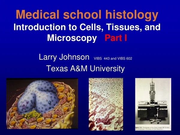
Medical school histologyIntroduction to Cells, Tissues, and Microscopy Part I Larry Johnson VIBS 443 and VIBS 602 Texas A&M University
OBJECTIVES Part I 1. Preview cellular ultrastructure • Preview cells, tissues, and organs Part II • Overview of light and electron microscopy 4. Preparation of specimens – types of visions 5. Section orientation
Introduction to HISTOLOGY PROTOPLASM – Living Substance ORGAN – Two or more types of tissues; larger functional unit e.g., skin, kidney, intestine, blood vessels CELL – Smallest unit of protoplasm Simplest animals consist of a single cell. CELL • TISSUE – Groups of cells with same general function and texture (texture = tissue) • e.g., muscle, nerve, epithelium, and connective tis. TISSUE ORGAN • ORGAN SYSTEM - Several organs • e.g., respiratory, digestive, reproductive systems SYSTEM
tissue organ cell organ system
In the 1660s, Robert Hooke looked through a primitive microscope at a thinly cut piece of cork. He saw a series of walled boxes that reminded him of the tiny rooms, or cellula, occupied by monks, and he coined the word "cell.“ Cells in a plant Although animal cells do not have cell walls like plants, cells are the “building blocks of life” of both.
Cells are composed of the nucleus and the surrounding cytoplasmic.
NUCLEUS HETEROCHROMATIN - CONDENSED CHROMTAIN EUCHROMATIN – DISPERSED FOR ACTIVE TRANSCRIPTION
NUCLEOLUS Pituitary CELL METABOLIC ACTIVITY OF CELL REFLECTED IN SIZE OF NUCLEOLUS • IF AXON IS CUT, SIZE OF NUCLEOLUS INCREASES BY 50% EGG SERTOLI CELL LEYDIG CELL NERVE CELL
CELL COMPOSITION THREE MAJOR CLASSES OF CYTOPLASMIC STRUCTURE -------------------------------------------------------------------------------- • MEMBRANOUS ORGANELLES - COMMON STRUCTURES, METABOLIC FUNCTION, CELL MEMBRANE, RER, SER, GOLGI, MITOCHONDRIA, LYSOSOMES • NON-MEMBRANOUS ORGANELLES – CYTOSKELETAL COMPONENTS, MICROTUBULES, MICROFILAMENTS, INTERMEDIATE FILAMENTS, FREE RIBOSOMES 3.INCLUSIONS - EXPENDABLES a. NUTRIENT e.g., GLYCOGEN, LIPID b. PIGMENT e.g., MELANIN GRANULES c. SECRETORY GRANULE e.g., ZYMOGEN GRANULE OF PANCREAS
Cytoplasm Pituitary CELL Metabolic activity of a cell is reflected in the amount of cytoplasm too. EGG SERTOLI CELL LEYDIG CELL NERVE CELL
Cellular ultrastructure EM 4c 60,000x EM 8f 9,000x Compare sizes of membranes ribosomes mitochondria EM 12a 13,200x
EM 6a 200,000x EM 2b 60,000x Compare sizes of membranes ribosomes mitochondria EM 7 80,000x
FOUR BASIC TYPES OF TISSUES IN THE BODY----------------------------------------------- Epithelium Connective tissue Muscular tissue Nervous tissue
Epithelium Functions: Cover organs, line viscera and blood vessels, secretory cells of glands
Epithelium Distinguishing features and distribution: • Always sit on a basement membrane, but come in a variety of configurations: classified on the basis of their shape and of the surface cells and whether one (simple) or more (stratified) layers of cells are stacked upon each other. • These cells are always attached to their neighbors by desmosomes, tight junctions, and gap junctions.
EpitheliumHistological Identification Simple Squamous – single layer of flat cells (blood vessels, covering of organs) Stratified Squamous – Multiple layers of cells with flat ones at the surface (skin, gums)
EpitheliumHistological Identification Simple cuboidal – Single layer of square cells (kidney tubules, liver cells, many others) Simple columnar – Single layer of tall, thin cells (intestinal epithelium)
EpitheliumHistological Identification Pseudostratified columnar – single layer of tall, thin cells packed together in such a jumble that they seem to be in layers, although all of the cells reach the basement membrane (respiratory passage) Transitional – stratified cuboidal epithelium of urinary passages
EpitheliumHistological Identification Some epithelia have surface specializations such as numerous microvilli or cilia.
EPITHELIUM CONNECTIVE TISSUE Skin Gunther von Hagens’ BODY WORLDS
Connective Tissue Function: the histological glue which binds the other tissues together to form organs, specializations include blood, cartilage, and bone. CONNECTIVE TISSUE
Connective Tissue Distinguishing features and histological identification: • Loose connective tissue – sparse collagen and elastic fibers, plentiful cells including fibroblasts, leukocytes • Dense connective tissue – concentrated collagen, few cells • Cartilage – avascular homogeneous matrix of collagen and protein-polysaccharides with few cells • Bone – calcified collagen matrix with few cells trapped in the caves of bone
Fat Muscle Gunther von Hagens’ Body Worlds
Muscle Function: generation of contractile force. Distinguishing features: • High concentration of contractile proteins actin and myosin arranged either diffusely in the cytoplasm (smooth muscle) or in regular repeating units called sarcomeres (striated muscles, e.g., cardiac and skeletal muscles).
Muscle Histological identification: • Skeletal muscle – very long cylindrical striated muscle cells with multiple peripheral nuclei • Cardiac muscle – short branching striated muscle cells with one or two centrally located nuclei • Smooth muscle – closely packed spindle-shaped cells with a single centrally placed nucleus and cytoplasm that appears homogeneous by light microscopy
Muscle Distribution: • Smooth – fusiform cells associated with the viscera, respiratory tract, blood vessels, uterus, etc. • Cardiac – striated muscles associated with the heart • Skeletal – striated muscles mostly associated with the skeleton
Smooth muscle NERVOUS TISSUE Gunther von Hagens’ Body Worlds
Nervous Tissue Functions: specialized for the transmission, reception, and integration of electrical impulses Distinguishing features: • neurons – very large excitable cells with long processes called axons and dendrites. The axons make contact with other neurons or muscle cells at a specialization called a synapse where the impulses are either electrically or chemically transmitted to other neurons or various target cells (e.g., muscle). Others secrete hormones.
Nervous Tissue Glial cells – the supporting cells of nervous tissue. Nerves – collections of neuronal processes bound together by connective tissue. Axons may be coated by a myelin sheath (“myelinated”) or simply protected by being cradled in an indentation of a glial cell (“unmyelinated”).
Nervous Tissue Distribution: • Comprise the central nervous system • Individual peripheral nerves are found throughout the body • Individual neurons and clusters of neurons (called ganglia) are found in most organs
Gunther von Hagens’ Body Worlds
Where are these basic tissues located? EPITHELIUM CONNECTIVE TISSUE MUSCULAR TISSUE NERVOUS TISSUE Epithelium
Where are these basic tissues located? EPITHELIUM CONNECTIVE TISSUE MUSCULAR TISSUE NERVOUS TISSUE Epithelium
Where are these basic tissues located? EPITHELIUM CONNECTIVE TISSUE MUSCULAR TISSUE NERVOUS TISSUE Connective tissue
Where are these basic tissues located? EPITHELIUM CONNECTIVE TISSUE MUSCULAR TISSUE NERVOUS TISSUE Muscular tissue
Where are these basic tissues located? EPITHELIUM CONNECTIVE TISSUE MUSCULAR TISSUE NERVOUS TISSUE NERVOUS TISSUE
Where are these basic tissues located? EPITHELIUM CONNECTIVE TISSUE MUSCULAR TISSUE NERVOUS TISSUE NERVOUS TISSUE
Conclusions • Preview cells and cellular ultrastructure • Preview tissues and organs
Medical school histologyIntroduction to Cells, Tissues, and Microscopy Part II Larry Johnson VIBS 443 and VIBS 602 Texas A & M University
OBJECTIVES 1. Preview cellular ultrastructure • Preview cells, tissues, and organs • Overview of light and electron microscopy 4. Preparation of specimens – types of visions 5. Section orientation
Magnification vs. Resolution • magnification - increase in image size • resolution - smallest distance between two points that can be seen (distinguished) Calculated by: 0.61 (wavelength) / numerical aperture 0.25 um for light microscope 0.1 nm for electron microscope
Sample Preparation • Fixation • Embedding A. Paraffin B. Plastic • Sectioning A. 0.5 um for Light Microscopy B. 60-80 nm for Electron Microscopy