Insights into HIV-1 gp120 V3 Loop Conformation and Antibody Interactions
340 likes | 471 Vues
This study examines the gp120 protein of HIV-1, focusing on the critical V3 loop region, which plays a significant role in antibody recognition and viral properties. The research builds upon findings from Stanfield et al. (2003) and emphasizes the importance of V3's structural variations in influencing infection and immune responses. By creating model structures for various Fabs, the work elucidates how these antibodies interact with distinct V3 conformations. Such insights are vital for understanding HIV-1 pathogenesis and developing effective therapies.

Insights into HIV-1 gp120 V3 Loop Conformation and Antibody Interactions
E N D
Presentation Transcript
Recurring conformation of the human immunodeficiency virus type 1 gp120 V3 loopRobyn L. Stanfield, Jayant B. Ghiara, Erica O. Saphire, Albert T. Profy, Ian A. Wilson Biol 368 Bobby Arnold Isaiah Castaneda
Outline • Human immunodefiency virus type 1 make-up • Overview of the Stanfield et. Al 2003 article • Importance of the V3 region • Creating model structures for different Fabs • Findings of the study
HIV-1 has an outer surface made up of glycoproteins • These proteins are named gp120 and gp41 • Result from cleavage of gp160 chain • They are noncovalently associated • Gp120 binds to CD4 • Contains 5 variable regions • They are known as V1-V5
Outline • Human immunodefiency virus type 1 make-up • Overview of the Stanfield et. Al 2003 article • Importance of the V3 region • Creating model structures for different Fabs • Findings of the study
The Stanfield et. al study looks closely at the V3 region • By doing so, they hope to learn • How the V3 loop is recognized by antibodies • How alterations affect the properties of the virus • Sequence • Conformation • Exposure
Outline • Human immunodefiency virus type 1 make-up • Overview of the Stanfield et. Al 2003 article • Importance of the V3 region • Creating model structures for different Fabs • Findings of the study
The V3 region is of particular interest • Variation in this region has been linked to several factors • change in cell types that get infected • Induction of syncitia • Ability to be neutralized • Progression to AIDS
Outline • Human immunodefiency virus type 1 make-up • Overview of the Stanfield et. Al 2003 article • Importance of the V3 region • Creating model structures for different Fabs • Findings of the study
Fabs are antibodies which bind to a V3 conformation • 4 Fabs - 59.1, 83.1, 50.1, 58.2 • Fab 83.1, 50.1, and 59.1 all bind to similar conformations of V3 • V3 loops are stabilized to prevent conformation change • 5 antibodies chosen from over 85,000 possible for their neutralizing and stabilizing qualities
Fab models are purified, crystallized, then evaluated • Antibodies isolated from mice • Fab fragments collected from antibodies, then purified • Fab mixed with 16-mer peptide MP1, and made into crystals on thin plates • Structures determined, then models created from Fab 58.2
Comparing V3 Peptide Conformations V3 peptides bound to Fabs50.1 & 59.1 Conformation is least similar to others, but still binds to neutralizing antibodies d. Type 1 turn conformation e. Distorted type 1 turn around GPGR f. Extended KRIHI region, like a. & b. Gamma-turn around GPGR g. Unclassified turn around GPGR
Outline • Human immunodefiency virus type 1 make-up • Overview of the Stanfield et. Al 2003 article • Importance of the V3 region • Creating model structures for different Fabs • Findings of the study
Analysis of structures showed higher R values • Refinement and rebuilding of structures caused high R values • Electron density maps were of good quality • Rfree 32.6%, Rcryst 28.8% where average for 2.6A is typically 25-29% for Rfree
The relatively large amount of weak data contributed to higher R values Strong data Weak data
Final statistics of the refined structure are shown in the table to the right • Number of reflections measured • out of reflections present at the • given resolution • Amount that atoms oscillate • By including # for different components, users can more easily detect error • Used in structure refinement • A well refined model should not have many plots in the disallowed region
Electron Density of V3 peptide bound to 83.1 Despite good quality maps, there was still room for refinement
Analyzing the molecular structure • Light and heavy chains categorized into L and H groups in table 3 • CDR portion of each molecule in bold • CDR - complementarity-determining region
Below are the sequences of the Fabs • This table will be referred to again when the issue of • kinks arises
All CDR loops normal except L1 • L1 CDR has 5 AA insertion after residue L27 • Tip of the loop bends away from antigen binding site • L1 loops move about 9 A from corresponding spot in other Fabs
Fab 83.1-V3 Complex • Light chains are in green • Heavy chains are in blue • V3 Peptide is in ball & stick model • Used MolScript program for the data
The L1 CDR loop of Fab 83.1bends away from the typical position • 27 other domains in grey • Fab 83.1 in red The bending is to avoid clashing with H41 & H42, shown in blue
CDR H3 shows a kinked base • Kinked base not predicted by sequence • Seen in at least 2 other Fabs, also not predicted • A salt bridge is usually formed with AspH101 and Arg/Lys, but does not for some reason
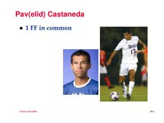
A straight torso was expected because there is no Arg • or Lys at H94 for the Asp to form a salt bridge with • This table will be referred to again when the issue of • sequence homology arises AlaIleAsp
The sequence indicated that H3 loops would have an extended torso • However, they are actually kinked
Peptide complex connect with light and heavy Fab chains • 110 total contact points • 7 Hydrogen bonds no charge-charge interaction • 6 Hydrogen bonds peptide-main chain • 1 bond to Arg side chain • H3 CDR most contacts
The table below shows all hydrogen bonds and the distance between them
Conformation of all but 58.2 similar • 58.2 varies around the residues at the tip of V3 • Do not show structural homology • Similarity in conformation is not due to similarity of Fabs • All 4 antibodies generated from similar mice
Comparing the V3 peptide-Fab complexes • Overall shape is similar • Extended regions • Crowns
Peptides bind in different locations and ways on antibody • Antibodies chosen for ability to neutralize, bind viruses • Conformations show preferred V3 loop • V3 structures shown show a recurring conformer on virus
This is how the V3 peptide binds with Fab 83.1 • Light chains labeled in red • Heavy chains labeled in blue Peptide makes 110 contacts Peptide makes 107 contacts
The peptides adopt the same shape, but bind in different orientations and locations • The antibodies are structurally and sequentially very different • Binding sites differ as well • Still, peptide conformations remain similar
Range of V3 conformation defined by X-ray snapshots • V3 region contacts CCR5 and CXCR4 during infection • To fully determine range, a more complete understanding of quaternary gp120/gp41 oligomers along with the role of V3 in protein assembly • This will allow for understanding how HIV-1 completes receptor binding and viral fusion
References • Stanfield et al. Recurring conformation of the human immunodeficiency virus type 1 gp120 V3 loop. Virology 315 (2003) p. 159-173. October 12 2011.