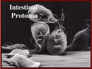
Intestinal` Protozoa
Intestinal` Protozoa. Giardia lamblia. Giardia trophozoites (light microscope). Giardia trophozoites ( SEM). Giardia trophozoites (light microscope). Giardia cyst (light microscope). Giardia trophozoites (light microscope). Trichrome stain. Unstained.

Intestinal` Protozoa
E N D
Presentation Transcript
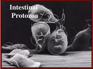
Giardia lamblia Giardia trophozoites (light microscope) Giardia trophozoites ( SEM)
Giardia trophozoites (light microscope)
Giardia cyst (light microscope)
Giardia trophozoites (light microscope) Trichrome stain Unstained
Giardia lamblia : Life cycle
Giardia cyst (light microscope) Trichrome stain Unstained
Giadriasis: Clinical Picture • Asymptomatic infections ( majority) • Symptomatic Infections: • Typical picture: IP 1-2 wks followed by diarrhoea for about 6 wks, • Atypical : Severe diarrhoea , malabsorption especially in children
Giardiasis: Laboratory diagnosis • Stools examination :cysts or trophozoits • Examination of duodenal contents : trophozoites
Giardiasis: Chemotherapy • Drug of choice: Metronidazole
Endolimax nana cyst Endolimax nana cyst
Endolimax nana trophozoite Endolimax nana cyst
E. polecki cyst • E. polecki trophozoite
Entamoeba coli(drawing) Entamoeba coli (Eosin)
Entamoeba coli ( trophozoite) Entamoeba coli ( trophozoite) Entamoeba coli ( trophozoite)
E. histolytica E. histolytica
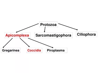
ENTAMOEBA HISTOLYTICA… 500 million people are infected. 100,000 deaths per year. Worldwide distribution. It is a waterborne infection. There are 6 species of Entamoeba: histolytica dispar hartmanni coli gingivalis polecki
E. histolytica vs E. dispar Entamoeba histolytica : amoebae that are pathogenic. E. dispar The non invasive form . The 2 amoebae can’t be distinguish by microscopic observation.
Entamoeba histolytica • Mode of infection • Water, food • Flies can act as vector.. • Can be sexually transmitted person -to -person contacts • Not a zoonosis
Entamoeba histolytica Life cycle Trophozoite: vegetative stage, must encyst to survive in the environment. It is a fragile structure. Pre cyst: encysting trophozoites disgorging any undigested food. Cyst: infective stage. Resist to the harsh conditions of the environment.
Entamoeba histolytica The infective dose can be as little as 1 cyst. be as little as 1 cyst. The incubation period can be from few days to can be from few days to few weeks depending on the infective dose Cysts can survive for weeks at appropriate weeks at appropriate temperature and humidity.
Entamoeba histolytica PATHOLOGY Intsetinal amoebiasis : Remarkable and unique ability a to hydrolyse host host tissues with their active cysteine proteases present on the surface membrane of the trophozoite. Lesions are found in the caecum, appendix, or colon. They may heal. If perforation of the colon occurs, They may heal. If perforation of the colon occurs, My lead to a peritonitis that can lead to death. Amoeboma Granuloma obstructing the bowel
PATHOLOGY : Intsetinal amoebiasis Complications
PATHOLOGY Intsetinal amoebiasis : Entamoeba histolytica
PATHOLOGY Intsetinal amoebiasis : Entamoeba histolytica
PATHOLOGY Intsetinal amoebiasis : E. Histolytica in mucosa. in mucosa. Numerous Numerous trophozoites can be seen with ingested seen with ingested erythrocytes.
Main Drugs for Treatment of Amoebiasis • Intestinal : • Asynpromatic (cysts only): diloxanide furoate (Furamide) • Sympromatic(cysts and trophozoites): metronidazole • Extra-intestinal: • Metronidazole
Free-living Amoebae: Naegleria fowleri and Acanthamoeba
Acanthamoeba keratitis • Patient upon presentation with Acanthamoeba keratitis. No severe pain was experienced by this patient, which made diagnosis difficult
Acanthamoeba keratitis • The same patient who had just developed a classic ring infiltrate