Download
1 / 60
630 likes | 1.24k Vues
Antigens/Antibodies. Chapters 4-6. Antigen Any substance that can bind to an antibody or T cell receptor. Immunogen Any substance that can elicit an immune response All immunogens are antigens Not all antigens are immunogens, i.e. haptens. Antigen vs. Immunogen. Determined by

E N D
Antigens/Antibodies Chapters 4-6
Antigen Any substance that can bind to an antibody or T cell receptor Immunogen Any substance that can elicit an immune response All immunogens are antigens Not all antigens are immunogens, i.e. haptens Antigen vs. Immunogen
Determined by Foreignness Molecular Size Chemical Composition Degradability Influenced by Host genotype Dose and route of administration Presence of adjuvants Immunogenicity of Antigens
Epitopes • Discrete sites on immunogens recognized by antibodies or T cell receptors • Macromolecules contain many epitopes with different immunogenicities • Immunogenicity can vary between individuals and populations. • Some epitopes are immunodominant
Identification of B-cell epitopes within the HPV16 E7 protein. Overlapping peptides were generated and injected into mice. The antibody response to each peptide was measured by ELISA. Comerford, S.A. et al. J. Virol. 65:4681-4690, 1991
Overlapping Non-overlapping Allosteric Linear Conformational Neoantigen Epitopes
B and T Cell Epitopes Can Overlap • Comerford, S.A. et al. J. Virol. 65:4681-4690, 1991
Objectives • To discuss the general properties of all immunoglobulins • To describe the basic structure of immunoglobulins • To relate immunoglobulin structure with function • To define immunoglobulin hypervariable and framework regions • To define immunoglobulin classes and subclasses, types and subtypes • To describe the structures and properties of immunoglobulin classes
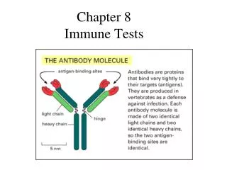
Immunoglobulin (Ig) • Immunoglobulinsare glycoprotein molecules that are produced by plasma cells in response to an immunogen and which function as antibodies. The immunoglobulins derive their name from the finding that they migrate with globular proteins when antibody-containing serum is placed in an electrical field. Electrophoretic separation of serum proteins
General Functions • Antigen Binding • Effector Functions
GENERAL FUNCTIONS OF IMMUNOGLOBULINS • Antigen binding • Immunoglobulins bind specifically to one or a few closely related antigens. • Binds to a specific antigenic determinant. • Antigen binding is the primary function of antibodies and can result in protection of the host. • The “valency” of antibody refers to the number of antigenic determinants that an individual antibody molecule can bind. • The valency of all antibodies is at least two and in some instances more.
Effector Functions • The immunoglobulins mediate a variety of these effectorfunctions: • Fixation of complement - This results in lysis of cells • Binding to various cell types: • Phagocytic cells, lymphocytes, platelets, mast cells, and basophils have receptors that bind immunoglobulins. This binding can activate the cells to perform some function. • Some immunoglobulins also bind to receptors on placental trophoblasts, which results in transfer of the immunoglobulin across the placenta. As a result, the transferred maternal antibodies provide immunity to the fetus and newborn
BASIC STRUCTURE OF IMMUNOGLOBULINS • Heavy and light chains • Disulphide bonds • Variable (V) and constant (C) regions • Hinge region • Domains • Oligosaccharides
IgG structure • 2 HEAVY chains =redandyellow • 2 LIGHT chains = green and blue. • 2 antiparallelbeta sheets • make up each of the immunoglobulin domains (2 Ig domains per L chain and 4 domains per H chain) • Papaincleavage site. • Papain cleavage gives ONE Fc fragment (2 immunoglobulin domains from each H chain) and • TWOFab fragments(each with 2 immunoglobulin domains from an H chain and 2 immunoglobulin domains from an L chain) • each Fab fragment contains ONE antigen binding site
plot of variability as a function of position along the sequence of the amino acids that make up the heavy chain of Ig molecules.
STRUCTURE OF THE VARIABLE REGION • Hypervariable (HVR) or complementarity determining regions (CDR) • Comparisons of the amino acid sequences of the variable regions of immunoglobulins show that most of the variability resides in three regions called the HVR/CDR. • Antibodies with different specificities (i.e. different combining sites) have different complementarity determining regions while antibodies of the exact same specificity have identical complementarity determining regions (i.e. CDR is the antibody combining site). • Complementarity determining regions are found in both the H and the L chains.
STRUCTURE OF THE VARIABLE REGION • Framework regions • The FR regions form a beta-sheet structure which serves as a scaffold to hold the HV regions in position to contact antigen. • Based on similarities and differences in the framework regions the immunoglobulin heavy and light chain variable regions can be divided into groups and subgroups. • Antibodies are divided into five major classes, IgM, IgG, Iga, IgD, and IgE, based on their framework/constant region structure.
HUMAN IMMUNOGLOBULIN CLASSES, SUBCLASSES, TYPES AND SUBTYPES • Immunoglobulin classesThe immunoglobulins can be divided into five different classes, based on differences in the amino acid sequences in the constant region of the heavy chains. • All immunoglobulins within a given class will have very similar heavy chain constant regions. These differences can be detected by sequence studies or more commonly by serological means (i.e. by the use of antibodies directed to these differences). • IgG - Gamma heavy chains • IgM - Mu heavy chains • IgA - Alpha heavy chains • IgD - Delta heavy chains • IgE - Epsilon heavy chains
Immunoglobulin Subclasses • The classes of immunoglobulins can de divided into subclasses based on small differences in the amino acid sequences in the constant region of the heavy chains. • All immunoglobulins within a subclass will have very similar heavy chain constant region amino acid sequences. Again these differences are most commonly detected by serological means. • IgG Subclasses • IgG1 - Gamma 1 heavy chains • IgG2 - Gamma 2 heavy chains • IgG3 - Gamma 3 heavy chains • IgG4 - Gamma 4 heavy chains • IgA Subclasses • IgA1 - Alpha 1 heavy chains • IgA2 - Alpha 2 heavy chains
IgG • Structure • All IgG's are monomers (7S immunoglobulin). The subclasses differ in the number of disulfide bonds and length of the hinge region.
Properties • IgG is the most versatile immunoglobulin because it is capable of carrying out all of the functions of immunoglobulin molecules. • IgG is the major Ig in serum - 75% of serum Ig is IgG • IgG is the major Ig in extra vascular spaces • Placental transfer - IgG is the only class of Ig that crosses the placenta. Transfer is mediated by a receptor on placental cells for the Fc region of IgG. Not all subclasses cross equally well; IgG2 does not cross well. • Fixes complement - Not all subclasses fix equally well; IgG4 does not fix complement • Binding to cells - Macrophages, monocytes, granulocytes and some lymphocytes have Fc receptors for the Fc region of IgG. Not all subclasses bind equally well; IgG2 and IgG4 do not bind to Fc receptors. A consequence of binding to the Fc receptors on PMNs, monocytes and macrophages is that the cell can now internalize the antigen better. The antibody has prepared the antigen for eating by the phagocytic cells. • IgG is a good opsonin.
IgG Increases in: • Chronic granulomatous infections • Infections of all types • Hyperimmunization • Liver disease • Malnutrition (severe) • Dysproteinemia • Disease associated with hypersensitivity granulomas, dermatologic disorders, and IgG myeloma • Rheumatoid arthritis Decreases in: • Agammaglobulinemia • Lymphoid aplasia • Selective IgG, IgA deficiency • IgA myeloma • Bence Jones proteinemia • Chronic lymphoblastic leukemia
Antibodies Continued
Reading • Chapter 4 Kuby et al, Immunology 6th Edition • Chapter 4 Abbas et al, Cellular and Molecular Immunology 6th Edition.
IgM • Structure: • Is a pentamer (19S Ig) but it can also exist as a monomer. • In the pentameric form all heavy chains are identical and all light chains are identical. • IgM has an extra domain on the mu chain (CH4) and it has another protein covalently bound via a S-S bond called the J chain. This chain functions in polymerization of the molecule into a pentamer.
Properties • Third most common serum Ig. • IgM is the first Ig to be made by the fetus and the first Ig to be made by a virgin B cells when it is stimulated by antigen. • Pentameric structure, IgM is a good complement fixing Ig - IgM antibodies are very efficient in leading to the lysis of microorganisms. • IgM is also a good agglutinating Ig - IgM antibodies are very good in clumping microorganisms for eventual elimination from the body. • IgM binds to some cells via Fc receptors.
Surface IgM Monomer No J chain Extra 20 aa in tail Associated with two additional proteins involved in signal transduction
IgA • Structure • Serum IgA is a monomer but IgA found in secretions is a dimer • When IgA exits as a dimer, a J chain is associated with it. • When IgA is found in secretions is also has another protein associated with it called the secretory piece or T piece • IgA is sometimes referred to as 11S immunoglobulin.
Unlike the remainder of the IgA which is made in the plasma cell, the secretory piece is made in epithelial cells and is added to the IgA as it passes into the secretions. The secretory piece helps IgA to be transported across mucosa and also protects it from degradation in the secretions.
Properties • IgA is the 2nd most common serum Ig. • IgA is the major class of Ig in secretions - tears, saliva, colostrum, mucus. Since it is found in secretions secretoryIgA is important in local (mucosal) immunity. • Normally IgA does not fix complement, unless aggregated. • IgA can binding to some cells - PMN's and some lymphocytes.
IgD • Structure • Exists only as a monomer. • Properties • IgD is found in low levels in serum; its role in serum uncertain. • IgD is primarily found on B cell surfaces where it functions as a receptor for antigen. • IgD on the surface of B cells has extra amino acids at C-terminal end for anchoring to the membrane. • It also associates with the Ig-alpha and Ig-beta chains. • IgD does not bind complement.
IgE • Structure • IgE exists as a monomer and has an extra domain in the constant region. • Properties • IgE is the least common serum Ig since it binds very tightly to Fc receptors on basophils and mast cells even before interacting with antigen. • Involved in allergic reactions - binding of the allergen to the IgE on the cells results in the release of various pharmacological mediators that result in allergic symptoms. • IgE also plays a role in parasitic helminth diseases. Since serum IgE levels rise in parasitic diseases, measuring IgE levels is helpful in diagnosing parasitic infections. Eosinophils have Fc receptors for IgE and binding of eosinophils to IgE-coated helminths results in killing of the parasite. • d) IgE does not fix complement.
Isotypes of Light Chain • Kappa (κ) and Lamda (λ), which are distinguished by their carboxy-terminal constant (C) regions. • An antibody molecule has either two κ light chains or two λ light chains, but never one of each. • In humans, about 60% of antibody molecules have κ light chains, and about 40% have λ light chains. • Changes in this ratio can occur in patients with monoclonal B cell tumors because the neoplastic clone produces antibody molecules with the same light chain. • In fact, the ratio of κ-bearing cells to λ-bearing cells is often used clinically in the diagnosis of B cell lymphomas. • In mice, κ-containing antibodies are about 10 times more abundant than λ-containing antibodies. • Unlike in heavy chain isotypes, there are no known differences in function between κ-containing antibodies and λ-containing antibodies.
Natural Distribution and Production of Antibodies • Found in biological fluids throughout the body • On surface of limited number of cells • B cells are the only cells that synthesize antibodies
Interesting facts • A healthy 70-kg adult human produces about 2g-3g of antibodies every day. • Almost two thirds of this is IgA, which is produced by activated B cells and plasma cells in the walls of the gastrointestinal and respiratory tracts and actively transported into the lumens. • The large amount of IgA produced reflects the large surface areas of these organs. • Antibodies that enter the circulation have limited half-lives.
After exposure to an antigen, much of the initial antibody response occurs in lymphoid tissues: • the spleen, • lymph nodes, • mucosal lymphoid tissues, • Secreted forms of antibodies accumulate in the plasma (the fluid portion of the blood), in mucosal secretions, and in the interstitial fluid of tissues. • Secreted antibodies often attach to the surface of other immune effector cells, such as mononuclear phagocytes, natural killer (NK) cells, and mast cells, which have specific receptors for binding antibody molecules.
Looking at Structure of Ig Molecules • Breakthrough came when researchers discovered that: • multiple myeloma patients produced biochemically identical antibody molecules • Led to an extremely powerful technique for producing monoclonal antibodies, described by Georges Köhler and Cesar Milstein in 1975. • They developed a method for immortalizing individual antibody-secreting cells from an immunized animal by producing "hybridomas," each of which secreted individual monoclonal antibodies of predetermined specificity.