Download
1 / 31
310 likes | 591 Vues
Approach to Failed Shoulder Stabilization. M Prud’homme-Foster AHD April 5 th , 2012. Glenohumeral joint is most common large joint to dislocate 11.2 per 100,000 person-years 90% traumatic dislocation: anterior This review will focus primarily on anterior instability. Pathoanatomy

E N D
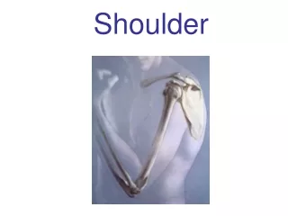
Approach to Failed Shoulder Stabilization M Prud’homme-Foster AHD April 5th, 2012
Glenohumeral joint is most common large joint to dislocate • 11.2 per 100,000 person-years • 90% traumatic dislocation: anterior This review will focus primarily on anterior instability Boone et al. Orth Clin of NA 2010 ; Shah et al. Artrhroscopy 2011 ; OKU 3 Shoulder & Elbow
Pathoanatomy • Clinical Evaluation • History and Evaluation • Imaging • Recurrent Instability • Surgical Treatment • Arthroscopic Bankart Revision • Open Bankart Revision • OsteoarticularPathalogy Boone et al. OrthClin of NA 2010 ; Shah et al. Artrhroscopy 2011 ; OKU 3 Shoulder & Elbow
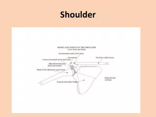
Pathoanatomy • GH relies on Dynamic and Static stabilizers • Critical static components • Glenolabral complex • Capsuloligamentous structures • Labrum increases surface area and depth: 50% Boone et al. OrthClin of NA 2010 ; Shah et al. Artrhroscopy 2011 ; OKU 3 Shoulder & Elbow
Boone et al. Orth Clin of NA 2010 ; Shah et al. Artrhroscopy 2011 ; OKU 3 Shoulder & Elbow
Pathoanatomy • Bankart lesion most common pathology with anterior instability • Anteroinferior labrum with attached IGHLC • Recurrent episodes—>elongation anteroinferior capsule • ‘Osseous Bankart’ Suguya 2003 • 50% of traumatic de novo and 86% in recurrent Boone et al. Orth Clin of NA 2010 ; Shah et al. Artrhroscopy 2011 ; OKU 3 Shoulder & Elbow
Cadaveric study with Bankart repair • Showed shoulder stable up to 21% defect • Instability with IR and restrictive ER
Bone loss affects contact pressures • Geiss et al. JSES 2002 • 30% glenoid defect increases contacts pressure 100% • Buscayret et al. Am J SpMed 2004 • Osseous defect and number of dislocation risk factor for OA Boone et al. Orth Clin of NA 2010 ; Shah et al. Artrhroscopy 2011 ; OKU 3 Shoulder & Elbow
Hill-Sachs lesion • Posterolateral aspect of head • Found in • 90% primary dislocators • 100% recurrent dislocators • 40% recurrent subluxators • Clinically significant threshold • 20-30% • Lesions parallel parallel to ant. glenoid surface in at risk position (Abd + ER) Boone et al. Orth Clin of NA 2010 ; Shah et al. Artrhroscopy 2011 ; OKU 3 Shoulder & Elbow
Clinical Evaluation Boone et al. Orth Clin of NA 2010 ; Shah et al. Artrhroscopy 2011 ; OKU 3 Shoulder & Elbow
History • Determine if instability and severity • Traumatic v. atraumatic • What was possibly overlooked? • 89% of failures have glenoid bone loss • Capsular laxity, HAGL, MDI, SLAP, Hill-Sachs, neuropraxia, SubS weakness, failed Bankart repair Boone et al. Orth Clin of NA 2010 ; Shah et al. Artrhroscopy 2011 ; OKU 3 Shoulder & Elbow
Physical Examination • Comparison to contralateral • Sulcus sign and Gagey signs: inferior laxity • A/PROM • ROM: chondrolysis, hardware impingement, overtightening of capsulolabral complex • RC evaluation • Higher incidence of RCT >40yoa • Open repair: 23% incompetent SubS (positive belly press) and 27% of contralateral • Apprehension and relocation testing Boone et al. Orth Clin of NA 2010 ; Shah et al. Artrhroscopy 2011 ; OKU 3 Shoulder & Elbow
Imaging • Radiographs: • AP, true AP, axillary • Westpoint • Anteroinferior bone loss • Styker Notch and AP in IR • Hill-Sachs Boone et al. Orth Clin of NA 2010 ; Shah et al. Artrhroscopy 2011 ; OKU 3 Shoulder & Elbow
CT Scan: Axial and sagittal views Itoi et al.: 21% represents 50% glenoid bone loss Saito et al.: 3 o’clock Boone et al. Orth Clin of NA 2010 ; Shah et al. Artrhroscopy 2011 ; OKU 3 Shoulder & Elbow
CT scan: humeral head subtracted • When length of lesion is half the diameter of the glenoid: 30% decrease in dislocation force
MR arthrography RCT, HAGL, capsular tears or laxity, posterior extension of Bankart MR not a substitute for CT
Recurrent Instability Boone et al. Orth Clin of NA 2010 ; Shah et al. Artrhroscopy 2011 ; OKU 3 Shoulder & Elbow
Prospective, 131 consecutive • Mean fu 31 months • Overall recurrence: • 15.3% • Recurrence with bone lesion or hyperlaxity • 75% • Open repair with more than 6 points • Similar studies support findings
Recurrent Instability: Closed v Open • Brophy 2009: roughly equal, 6.4% v 8.2% • Tauber 2004 at revision: 56% bony Bankart, 22% large capsule, 5% laterally torn capsule • Burkhart 2000: closed, 4% without bony, 67% with signicant • Yiannakopoulous 2007: 15.4% with inverted pear (28.8% loss) Boone et al. Orth Clin of NA 2010 ; Shah et al. Artrhroscopy 2011 ; OKU 3 Shoulder & Elbow
Recurrence: soft tissue • 83-91% reported to have poor capsulolabral tissue or redundancy • Asymmetric or overtightening: Erlenmeyer flask phenomena (tight anteriorly and superiorly and inferior instability)—22% • Improper position of anchors: @ margin of articular surface—46-100% Boone et al. Orth Clin of NA 2010 ; Shah et al. Artrhroscopy 2011 ; OKU 3 Shoulder & Elbow
Hawkins and Hawkins 1985: 38.7% due to misdiagnosis Boone et al. Orth Clin of NA 2010 ; Shah et al. Artrhroscopy 2011 ; OKU 3 Shoulder & Elbow
Boone et al. Orth Clin of NA 2010 ; Shah et al. Artrhroscopy 2011 ; OKU 3 Shoulder & Elbow
Surgery for revision Open Arthroscopic • Levine 2000: 17% failure or 44% if multiple revisions • Scar tissue affects abitility to maintain • Notable decrease in outcome and patient satisfaction • Careful selection: minimal bone loss <15% • Can identify ST pathologies and minimize iatrogenic (Subscap) • Limited evidence, 10-27% recurrence with 78% good-excellent results Boone et al. Orth Clin of NA 2010 ; Shah et al. Artrhroscopy 2011 ; OKU 3 Shoulder & Elbow
Osteoarticular: Laterjet or Bristow Boone et al. Orth Clin of NA 2010 ; Shah et al. Artrhroscopy 2011 ; OKU 3 Shoulder & Elbow
Subscapularis: to split or not? Positioning • L-shaped incision: 2/3 split • Decrease muscle power • L-shaped:42% • Split:8% • Lateralization: early OA • Mediliazation: instability • Capsular repair can make the corocoid intra or extra articular • Laterjet: 95% success (Burkart) Boone et al. Orth Clin of NA 2010 ; Shah et al. Artrhroscopy 2011 ; OKU 3 Shoulder & Elbow
Humeral Head defects • Important for 30% of articular surface • Structural allograft: sized matched humeral heads or femoral head • Artrhoplasty in older patients Boone et al. Orth Clin of NA 2010 ; Shah et al. Artrhroscopy 2011 ; OKU 3 Shoulder & Elbow
Capsular plication Boone et al. Orth Clin of NA 2010 ; Shah et al. Artrhroscopy 2011 ; OKU 3 Shoulder & Elbow
Boone et al. Orth Clin of NA 2010 ; Shah et al. Artrhroscopy 2011 ; OKU 3 Shoulder & Elbow