Knee Anatomy
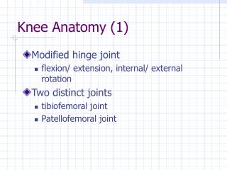
Knee Anatomy. Sports Medicine 2 J. Cresimore EFHS. Knee Joint. The most poorly constructed joint in the body. Femur round, tibia flat. Comprised of four bones. Femur Tibia Fibula Patella. Femur. Medial and Lateral Condyles- distal ends of the femur. Patella.


Knee Anatomy
E N D
Presentation Transcript
Knee Anatomy Sports Medicine 2 J. Cresimore EFHS
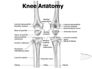
Knee Joint • The most poorly constructed joint in the body. Femur round, tibia flat. • Comprised of four bones. • Femur • Tibia • Fibula • Patella
Femur • Medial and Lateral Condyles- distal ends of the femur.
Patella • Patella tendon- attaches to the anterior of the tibia. • Quadriceps tendon-attaches the quadriceps to the patella.
Cruciate Ligaments • Major stabilizing ligaments in the knee • Anterior Cruciate Ligament (ACL)-prevents the tibia from sliding out in front of the femur • Injuries caused by hyperflexion
Cruciate Ligaments • Posterior Cruciate Ligament (PCL)-It prevents the tibia from sliding backwards under the femur. • Injuries usually caused by Hyperextension
Collateral Ligament • Medial Collateral Ligament (MCL)- connect the tibia and the femur. • A force from the lateral side could cause a tear.
Collateral Ligament • Lateral Collateral Ligament (LCL)- connect the fibula to the femur. • A force from the medial side can cause a tear of the LCL
How are ligaments torn? • Medial collateral ligament (MCL) is injured from a blow/force to the outside of the leg. • Lateral collateral ligaments are torn blow/force to the inside of the leg.
Cartilage • Articulate Cartilage-covers the moving parts of the knee. • Chronic damage to articulate cartilage leads to arthritis.
Cartilage • Meniscus- half moon shaped cartilage lying between the knee joint.