

SlideServe
Menu
Préférences
© SlideServe · v2026

Knee Anatomy

adolph avatar
30 diapositives · 2.8K Views · Oct 09, 2014

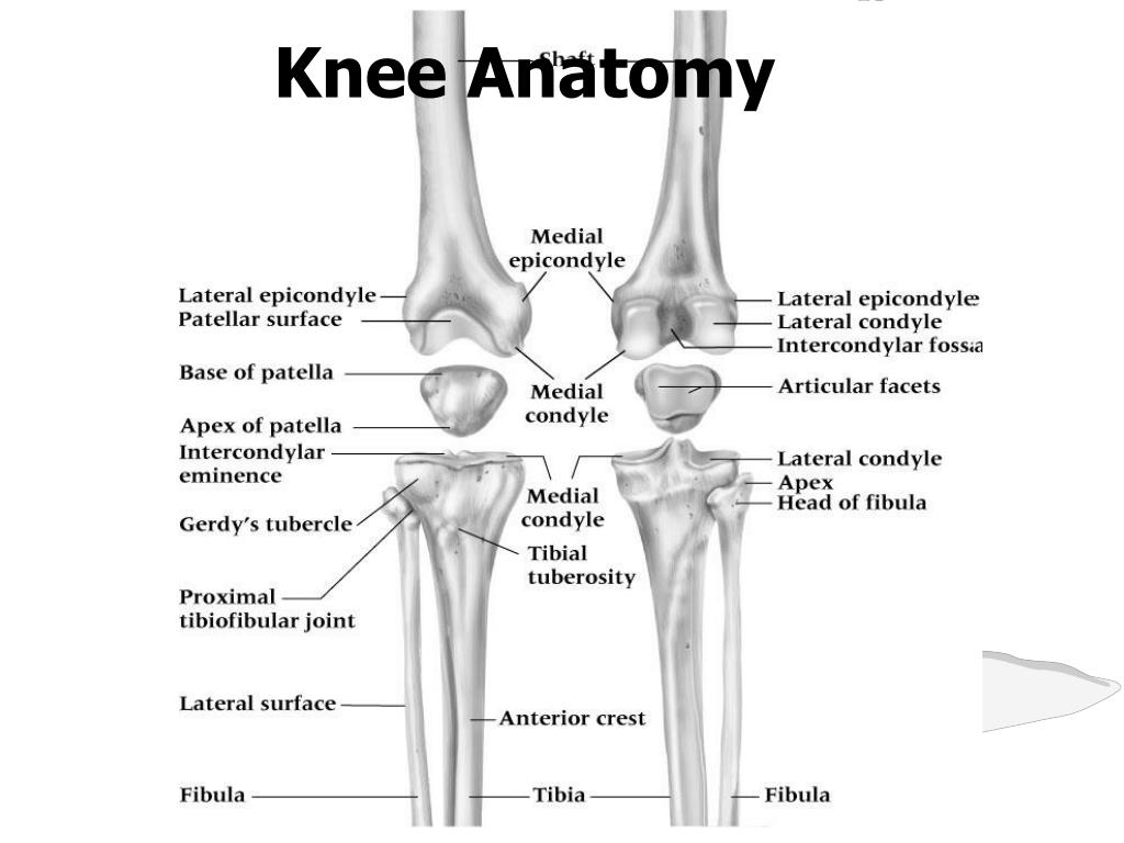
Knee Anatomy. Knee Joint. The most poorly constructed joint in the body. Femur round, tibia flat. Comprised of four bones. Femur Tibia Fibula Patella. Femur. Medial and Lateral Condyles- distal ends of the femur. Largest bone in the body. Femur. Landmarks to know Add. Tubercle
Knee Anatomy - slide 1
01 / 30Knee Anatomy
1 / 17

Télécharger la présentation

Knee Anatomy
Une image / un lien ci-dessous est fourni (tel quel) pour télécharger la présentation

Politique de téléchargement : le contenu du site vous est fourni TEL QUEL pour votre information et votre usage personnel ; il ne peut être vendu, concédé sous licence ni partagé sur d’autres sites sans l’accord de son auteur.Le contenu vous est fourni TEL QUEL, pour votre information et votre usage personnel uniquement. Téléchargez la présentation en cliquant sur ce lien. Lors du téléchargement, si vous ne parvenez pas à télécharger une présentation, l’éditeur a peut-être supprimé le fichier de son serveur.Pendant le téléchargement, si vous ne parvenez pas à obtenir une présentation, le fichier a peut-être été supprimé par l’éditeur.

F I N

Plus de contenu associé

Transcription de la présentation
  1. Knee Anatomy

  2. Knee Joint • The most poorly constructed joint in the body. Femur round, tibia flat. • Comprised of four bones. • Femur • Tibia • Fibula • Patella

  3. Femur • Medial and Lateral Condyles- distal ends of the femur. • Largest bone in the body

  4. Femur • Landmarks to know • Add. Tubercle • Medial and Lateral epicondyles • Medial and lateral condyles • Intercondylar fossa • Patella fossa (not shown)

  5. Tibia and Fibula

  6. Fibula • Landmarks to know • Apex • Head • Neck • Lateral Maelleolus

  7. Tibia • Landmarks to know • Intercondylar eminence • Medial and lateral condyles • Tibial tuberosity

  8. Patella • Patella tendon- attaches to the anterior of the tibia. (tibial tuberosity) • Quadriceps tendon-attaches the quadriceps to the patella.

  9. Joints • Tibiofemoral • Largest joint in body • Patellofemoral • Patella contains the thickest cartilage found in the body • Superior Tibiofibular • Any movement here is due to movement at the ankle

  10. PRIMARY Medial Collateral Ligament Lateral Collateral Ligament Anterior Cruciate Ligament Posterior Cruciate Ligament SECONDARY Medial Meniscus “C” shaped Lateral Meniscus “O” shaped Knee Stabilizers

  11. Cruciate Ligaments • Major stabilizing ligaments in the knee • Anterior Cruciate Ligament (ACL)-prevents the tibia from sliding out in front of the femur • Injuries caused by hyperflexion, internal rotation, hyperextension

  12. ACL • Has Two Bundles • Anteromedial • Tight in flexion and extension • Posterolateral • Tight in extension • Ligament is most lax between 30 – 60 degrees flexion

  13. Posterior Cruciate Ligament • Prevents posterior translation of the tibia on the femur • Resists hyperextension of knee • Runs from posterior tibia to anterior femur

  14. PCL • Fibers are tightest around 30 degrees flexion • Posterolateral fibers are the last to become tight • Two times stronger than ACL

  15. Collateral Ligament • Medial Collateral Ligament (MCL)- connect the tibia and the femur. • A force from the lateral side could cause a tear. • Valgus force

  16. Medial Collateral Ligament • Two layers • Deep layer is actually a thickening of the joint capsule that blends into the medial meniscus • Superficial layer is what we view as the MCL

  17. Collateral Ligament • Lateral Collateral Ligament (LCL)- connect the fibula to the femur. • A force from the medial side can cause a tear of the LCL • Varus force

  18. Lateral Collateral Ligament • Attaches to head of fibula • Prevents excessive varus and IR forces • Tightest in extension, loosest after 30 degrees flexion

  19. Cartilage • Articulate Cartilage-covers the moving parts of the knee. • Chronic damage to articulate cartilage leads to arthritis.

  20. Cartilage • Meniscus- half moon shaped cartilage lying between the knee joint.

  21. ARTICULAR DISCS • Medial Meniscus • Lateral Meniscus

  22. Meniscal Blood Supply • Each Meniscus has 3 zones • Red Zone • Outer 1/3: good blood supply • Red/White Zone • Middle 1/3: minimal blood supply • White Zone • Inner 1/3: avascular (no blood supply) • Implications for injury?

  23. Meniscal Blood Supply