

SlideServe
Menu
Préférences
© SlideServe · v2026

Knee Anatomy

drago avatar
13 diapositives · 707 Views · Oct 09, 2014

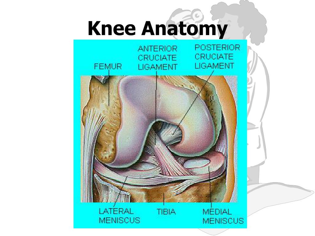
Knee Anatomy. Knee Joint. The most poorly constructed joint in the body. Femur has rounded distal epiphysis while proximal epiphysis of Tibia is flat. Creates a very unstable design. Comprised of four separate bones. Femur Tibia Fibula Patella. Femur and Tibia.
Knee Anatomy - slide 1
01 / 13Knee Anatomy
1 / 17

Télécharger la présentation

Knee Anatomy
Une image / un lien ci-dessous est fourni (tel quel) pour télécharger la présentation

Politique de téléchargement : le contenu du site vous est fourni TEL QUEL pour votre information et votre usage personnel ; il ne peut être vendu, concédé sous licence ni partagé sur d’autres sites sans l’accord de son auteur.Le contenu vous est fourni TEL QUEL, pour votre information et votre usage personnel uniquement. Téléchargez la présentation en cliquant sur ce lien. Lors du téléchargement, si vous ne parvenez pas à télécharger une présentation, l’éditeur a peut-être supprimé le fichier de son serveur.Pendant le téléchargement, si vous ne parvenez pas à obtenir une présentation, le fichier a peut-être été supprimé par l’éditeur.

F I N

Plus de contenu associé

Transcription de la présentation
  1. Knee Anatomy

  2. Knee Joint • The most poorly constructed joint in the body. Femur has rounded distal epiphysis while proximal epiphysis of Tibia is flat. Creates a very unstable design. • Comprised of four separate bones. • Femur • Tibia • Fibula • Patella

  3. Femur and Tibia • Articular cartilage covers the ends of both the femur and the tibia. • Medial and Lateral Condyles- found at the distal end of the Femur. • Medial and lateral Condyles found at the proximal end of the Tibia.

  4. Patella • Patella tendon- attaches onto the anterior of the Tibia. • Provides protection of Knee Joint, Patella also lifts tendons to increase leverage. • Quadriceps tendon-attaches the Quadriceps to the patella.

  5. Cruciate Ligaments • Major stabilizing ligaments in the knee. • Anterior Cruciate Ligament (ACL)-prevents the tibia from sliding out in front of the femur. • Posterior Cruciate Ligament (PCL) prevents knee from hyperextending • Injuries are most often caused by hyperflexion and hyperextension of the knee or rotation at the knee.

  6. Cruciate Ligaments • Posterior Cruciate Ligament (PCL)-It prevents the tibia from sliding backwards under the femur. • Injuries usually caused by Hyperextension • These cruciate ligaments get their name for their cross over (crucifix) arrangement.

  7. Collateral Ligament • Medial Collateral Ligament (MCL)- connect the tibia and the femur on the medial side of the knee joint. • A force from the lateral side could cause a tear to MCL. • Very common injury in many contact sports.

  8. Collateral Ligament • Lateral Collateral Ligament (LCL)- connect the fibula to the femur on the lateral side of knee. • A force from the medial side (rare) can cause a tear of the LCL.

  9. Cartilage • Articulate Cartilage-covers the moving parts of the knee. • Very smooth and slippery. • Chronic damage to articulate cartilage leads to osteoarthritis.

  10. Cartilage • Meniscus- half moon shaped cartilage (Fibrocartilage) lying between the knee joint. • These Menisces (Lateral/Medial) act as shock absorbers to handle pounding in the joint. • Often prone to tearing, which may require surgery to remove fragment.

  11. Meniscus Tear

  12. Knee Injuries • ACL Replacement surgery. • Often requires harvesting 1/3rd of Patellar tendon or from cadaver. • Animation • Animation 2

  13. Arthroscopic Surgery for Torn Meniscus http://www.youtube.com/watch?v=pguNCtOwzEc http://www.youtube.com/watch?v=i8EpT3uCVWU Checkout Animation on Arthroscopic Surgery KNEE SURGERY