Download
1 / 29
290 likes | 498 Vues
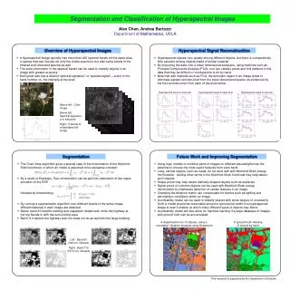
SEGMENTATION OF ARTERIAL GEOMETRY FROM ULTRASOUND IMAGES USING BALLOON MODELS. Author: Chaopquan Chen, Tamie L Poeppin Jason J Beech-Brandt etc. Source: IEEE International Symposium 2004 Page(s):1319 - 1322 Vol. 2 Student: Jia – Hong Chen Advisor: Ku – Yaw Chang. OUTLINE.

E N D
SEGMENTATION OF ARTERIAL GEOMETRY FROM ULTRASOUND IMAGES USING BALLOON MODELS Author: Chaopquan Chen, Tamie L Poeppin Jason J Beech-Brandt etc. Source: IEEE International Symposium 2004 Page(s):1319 - 1322 Vol. 2 Student: Jia – Hong Chen Advisor: Ku – Yaw Chang
OUTLINE • INTRODUCTION • MODEL DESCRIPTION • EXPERIMENTAL VALIDATION • 3D SEGMENTAION AND MESHING • CONCLUSIONS
INTRODUCTION • Cardiovascular disease accounts for 50% of all world deaths. • Traditional imaging methods • MRI • Ultrasound • Segmentation of Arterial geometry • B-mode ultrasound • MRI • spiral angiography
INTRODUCTION • 超音波系統通常依其所採用顯影型態分類,主要分為三大類 • A-模超音波(A-mode ultasound) • B-模超音波(B-mode ultasound) • 都卜勒
INTRODUCTION • A-模超音波(A-mode ultasound) • 將回音訊號以波形顯示,僅能顯示反射波的強度與組織介面的深度。通常用於船隻偵測海底深度。 • B-模超音波(B-mode ultasound) • 將回音訊號以光點顯示,可呈現組織的平面動態影像。Ex.懷孕時照的腹部超音波即為此類型。
INTRODUCTION • 都卜勒 • 連續波都卜勒: 超音波訊息不斷的放出和接收,可偵測範圍很廣的血流速度。 • 脈搏波都卜勒: 可定位某一深度與範圍血流方向速度,無法顯示影像,一般傳統穿顱超音波即使用此方法。 • 複合都卜勒超音波: 同時將B-模超音波與脈搏波都卜勒結合在一起,可同時顯示組織與血管的構造及血管管腔內的血液方向與流速。 • 彩色都卜勒超音波: 將都卜勒超音波顯示的血流方向賦予不同顏色,如朝向探頭的血流使用紅色,遠離探頭的血液使用藍色,亂流用綠色等。頸動脈超音波與顱內超音波即使用彩色都卜勒超音波。 • 動力都卜勒超音波: 以顏色顯示訊號的強度而非流速,無方向性,但不受超音波入色角度影響,不會產生錯像(aliasing),可偵測很低流速的訊號。
INTRODUCTION • Ultrasound has a number of features which will make segmentation challenging: • Speckle • Directionality • Data loss
INTRODUCTION • Speckle • interference of acoustic wavelets • not dependent on signal intensity • Directionality • loss of information at the lateral surfaces • Data loss • Calcification may cause shadowing of deeper structures
INTRODUCTION • Deformable models • extensively used in medical image processing and analysis • particularly to locate object boundaries • global information is pursued with deformable models • Snake • balloon models • the major difficulties • catching range’ • effect of image features
OUTLINE • INTRODUCTION • MODEL DESCRIPTION • EXPERIMENTAL VALIDATION • 3D SEGMENTAION AND MESHING • CONCLUSIONS
MODEL DESCRIPTION • Snake(active contours) • Energy-minimizing parametric curves • Internal stretching force and bending force • Pressure force • Closed models inflate like balloons
MODEL DESCRIPTION 內部能量的拉伸和彎曲 圖像本身的能量
MODEL DESCRIPTION 考慮直線:做一階微分(一~三點) 考慮曲線:做二階微分(多點) 二維高斯函數 Laplacian operator 影像強度
MODEL DESCRIPTION • Gaussian function • Catching area of the image feature • Reduce speckle • In B mode ultrasound images • tissue and blood can be characterized by mean intensity m and standard deviation σ
MODEL DESCRIPTION 平均強度 標準差
MODEL DESCRIPTION Figure 1 Image force defined by combining the gradient and the second order derivative. It disappears in the location of the desired edge and pushes towards each other from the inside and outside.
MODEL DESCRIPTION • Two problem will be encountered with traditional snake models • Cure initialized is not close enough to the boundary, it will not be driven to desired position • if the curve is not subjected to any forces, it will tend to shrink • Therefore, the image force is modified by an additional pressure force
MODEL DESCRIPTION 單位法向量 增加強度
MODEL DESCRIPTION Figure 2 ‘Pressure’ direction decided by examining the intensity and its change along the normal in (a). The contour will be driven toward the desired boundary from both directions in (b).
OUTLINE • INTRODUCTION • MODEL DESCRIPTION • EXPERIMENTAL VALIDATION • 3D SEGMENTAION AND MESHING • CONCLUSIONS
EXPERIMENTAL VALIDATION A:描述輪廓的形狀 B:描述所對應的黃金比例
EXPERIMENTAL VALIDATION Figure 3 Examples of artery images where lumen boundary were found by the model
OUTLINE • INTRODUCTION • MODEL DESCRIPTION • EXPERIMENTAL VALIDATION • 3D SEGMENTAION AND MESHING • CONCLUSIONS
3D SEGMENTAION AND MESHING • A 3D dataset consists of a set of 2D slices obtained • sweeping of the transducer along the surface of thepatients’ skin or the surface of a flow model.
3D SEGMENTAION AND MESHING Figure 5 3D mesh of a phantom artery segmented by the model
OUTLINE • INTRODUCTION • MODEL DESCRIPTION • EXPERIMENTAL VALIDATION • 3D SEGMENTAION AND MESHING • CONCLUSIONS
Conclusion • A modified balloon was built to segment arterial geometryfrom ultrasound images. • Theexperiment shows the accuracy of the model can bewithin 0.40±0.30mm. • This can provide thegeometry for the input into blood and tissue modeling
THE END Thanks~