MALE REPRODUCTIVE SYSTEM
780 likes | 1.17k Vues
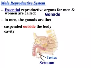
MALE REPRODUCTIVE SYSTEM. MALE REPRODUCTIVE SYSTEM. TO REVIEW THE COMPONENTS OF THE MALE REPRODUCTIVE SYSTEM. TO CHARACTERIZE THE GENERAL ORGANIZATION OF THE TESTIS. TO UNDERSTAND THE HORMONAL REGULATION AND PROCESS OF SPERMATOGENESIS. MALE REPRODUCTIVE SYSTEM. TESTES.

MALE REPRODUCTIVE SYSTEM
E N D
Presentation Transcript
MALE REPRODUCTIVE SYSTEM TO REVIEW THE COMPONENTS OF THE MALE REPRODUCTIVE SYSTEM TO CHARACTERIZE THE GENERAL ORGANIZATION OF THE TESTIS TO UNDERSTAND THE HORMONAL REGULATION AND PROCESS OF SPERMATOGENESIS
MALE REPRODUCTIVE SYSTEM TESTES EPIDIDYMIS VAS DEFERENS SEMINAL VESICLES PROSTATE BULBOURETHRAL GLANDS URETHRA
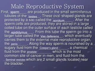
Male Reproductive System • Pathway of spermatozoa • Epididymis • Ductus deferens (Vas deferens) • Ejaculatory duct • Accessory organs • Seminal vesicles • Prostate gland • Bulbourethral glands • Scrotal sac encloses testes • Penis
The Male Reproductive System in Midsagital View Figure 28.1
The Male Reproductive System in Anterior View Figure 28.3
The Structure of the Testes Figure 28.4
The Epididymus Figure 28.9
Epididymis and Ductus Deferens • Epididymis: connection between thetestisand ductus deferens • Three parts: head (cauda), body, and tail • Ductus deferens(vas deferens): connects tail of epididymis to ejaculatory duct • Ascends within scrotum in the spermatic cord, into the pelvic region.
The Ductus Deferens and Accessory Glands Figure 28.10a-e
Seminal Vesicles and Prostate • Seminal vesicles: enlarged, sac-like structures which open into ejaculatory ducts. • Prostate: lobular structure at base of bladder.
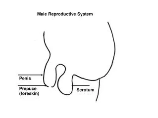
Male External Genitalia • Penis: male copulatory organ (also excretion: elimination of urine) • Composed of three columns of erectile tissue, each enclosed by connective tissue • Scrotum: two separate compartments, each containing a testis. • Wall of scrotum: skin, superficial loose connective tissue, smooth muscle (dartos).
Male Accessory Sex Organs: the Penis • 3 columns of erectile tissue: - corpus spongiosum: 1 column, includes glans penis, and contains the spongy (penile) urethra - 2 corpora cavernosa: lateral columns, forming the dorsum and sides of the penis
The Penis Figure 28.11
Hormones and Male Reproductive Function • FSH (Follicle stimulating hormone) • Targets sustentacular cells to promote spermatogenesis • LH (leutinizing hormone) • Causes secretion of testosterone and other androgens • GnRH (Gonadotropin releasing hormone) • Testosterone • Most important androgen
MALE REPRODUCTIVE SYSTEM TESTIS TUNICA ALBUGINEA - thick connective tissue capsule - connective tissue septa divide testis into 250 lobules - each lobule contains 1-4 seminiferous tubules and interstitial connective tissue (1) SEMINIFEROUS TUBULES - produce sperm INTERSTITIAL TISSUE - contains Leydig cells which produce testosterone (2) RECTUS TUBULES (3) RETE TESTIS (4) EFFERENT DUCTULES (5) EPIDIDYMIS
MALE REPRODUCTIVE SYSTEM TUNICA ALBUGINEA TESTIS Mediastinum containing RETE TESTIS EPIDIDYMIS LOBULES
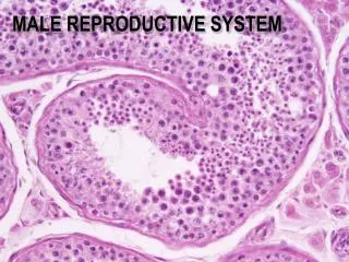
TESTIS H&E SEMINIFEROUS TUBULES
TESTIS H&E SEMINIFEROUS TUBULES SEMINIFEROUS TUBULES INTERSTITIAL CONN. TISSUE
TESTIS H&E SEMINIFEROUS TUBULES SEMINIFEROUS TUBULES INTERSTITIAL CONN. TISSUE
MALE REPRODUCTIVE SYSTEM TESTIS TUNICA VAGINALIS TUNICA ALBUGINEA SEMINIFEROUS TUBULES SEMINIFEROUS EPITHELIUM - complex stratified epithelium containing two basic cell populations: (1) SPERMATOGENIC CELLS (2) SERTOLI CELLS
MALE REPRODUCTIVE SYSTEM TESTIS SEMINIFEROUS TUBULES SEMINIFEROUS EPITHELIUM - complex stratified epithelium containing two basic cell populations: (1) SPERMATOGENIC CELLS stem cells which regularly replicate and differentiate into mature sperm as they migrate toward the lumen (2) SERTOLI CELLS nonreplicating physical support cells INTERSTITIAL CONNECTIVE TISSUE (1) LEYDIG CELLS produce and release testosterone
MALE REPRODUCTIVE SYSTEM SPERMATOGENESIS SPERMATOGONIA 1º SPERMATOCYTE 2º SPERMATOCYTE SPERMATIDS SPERMATIDS 2º SPERMATOCYTE 1º SPERMATOCYTE SERTOLI CELLS: - columnar with adjoining lateral processes - extend from basal lamina to lumen - Sertoli-Sertoli junctions divide seminiferous tubules into basal and adluminal compartments SERTOLI CELLS SPERMATOGONIA
- diploid cells (2n) created in spermatogonial phase give rise to haploid cells (1n) - Meiosis I (reduction division) and Meiosis II (equatorial division) - 1º spermatocytes enter Meiosis I to form 2º spermatocytes which then enter Meiosis II and result in spermatids MALE REPRODUCTIVE SYSTEM SEMINIFEROUS TUBULES SPERMATOGENESIS THREE PHASES: (1) Spermatogonial Phase (Mitosis) - spermatogonia proliferate by mitotic divisions to provide stem cells and cells which will proceed through spermatogenesis (1º spermatocytes) (2) Spermatocyte Phase (Meiosis) (3) Spermatid Phase (Spermiogenesis) - spermatid differentiation into spermatazoa
Basal Lamina Daughter cell Type A spermatogonium remain at basal lamina as a precursor cell 2n 2n Spermatogonia (stem cells) 2n mitosis Daughter cell Type B Spermatagonium Moves to adluminal compartment n 1° spermatocyte Meiosis I completed n 2° spermatocyte n Meiosis II n n n n Early spermatids n n n n Late spermatids
MALE REPRODUCTIVE SYSTEM SPERMATOGENESIS THREE PHASES: (1) Spermatogonial Phase (Mitosis) (2) Spermatocyte Phase (Meiosis) (3) Spermatid Phase (Spermiogenesis) - acrosome formation; golgi granules fuse to form acrosome that contains hydrolytic enzymes which will enable the spermatozoa to move through the investing layers of the oocyte - flagellum formation; centrioles and associate axoneme (arrangement of microtubules in cilia) - changes in size and shape of nucleus; chromatin condenses and shedding of residual body (cytoplasm)
MALE REPRODUCTIVE SYSTEM SPERMIOGENESIS Mature sperm 60µm long and acquire full motility in epididymis (1) HEAD - nucleus and acrosome (2) NECK - centriole and connecting piece (3) TAIL - middle piece (axoneme, outer dense fibers, mitochondial sheath) - principal piece (axoneme, outer dense fibers, fibrous sheath) - end piece (axoneme)
MALE REPRODUCTIVE SYSTEM SPERMIOGENESIS
Maturation of Sperm • Sperm leaving the testis and entering the epididymis are nonmotile and not capable of fertilization. • Sperm acquire motility and final maturation as they travel through the epididymis.
Time Span of Sperm Development • It takes about 70 days to develop from spermatogonia to spermatozoa. • It takes another 14 days to travel through epididymis to the ejaculatory duct. • Illness and exposure to toxic agents can have a delayed effect on quality of sperm produced.
Production of Semen • Semen is composed of spermatozoa and the secretory products of the seminal vesicle (60%) and prostate (30%). • Products of the seminal vesicle and prostate include - fructose (metabolized for energy by sperm) - prostaglandins (uterine contractions) - coagulating and decoagulating factors - antibacterial agents - pH adjusters (acids and bases) • The bulbourethral gland secretes an alkaline mucus, neutralizes acidity in the urethra and provides lubrication
Capacitation of Sperm • To fertilize an egg, the spermatozoa must undergo capacitation in the female reproductive tract following ejaculation. • Capacitation results in: - increased velocity of sperm movement - release of enzymes which allow sperm to reach the oocyte and penetrate it (acrosome reaction) - requires 2 to 6 hours (sperm may remain alive in the reproductive tract for days)
Male Sexual Response • Three distinct phases have been identified; arousal, emission, and ejaculation. • Arousal: erotic thoughts or physical stimulation result in activation of the parasympathetic system (via pelvic splanchnic nerves). - increased production of nitric oxide - nitric oxide activates soluble guanylate cyclase, resulting in increased production of cGMP - cGMP causes vasodilatation of blood vessels in the penis, resulting in increased blood flow and erection
Male Sexual Response • Emission: Sympathetic stimulation causes peristaltic contractions of the ampulla of the ductus deferens, the seminal vesicles, and the prostate. Thus, spermatozoa and seminal fluids enter the prostatic urethra. • At the same time, the internal urethral sphincter closes off the bladder to prevent retrograde ejaculation
Male Sexual Response • Ejaculation: Contractions of two skeletal muscles: - ischiocavernosus: contractions against the erectile tissue of the penis - bulbocavernosus: contractions push semen from base of penis to urethral opening (note that these are skeletal muscles under sympathetic control)
Role of Testosterone in Male Sexual Response • Testosterone increases libido (sexual thoughts and desires). • However, testosterone plays little role in capacity of men to have sexual intercourse. • Impotence (inability to achieve or maintain erection) can be due to physical causes (circulatory problems, drugs, alcohol, trauma, illness) or psychological state (depression, anxiety, stress). • Viagra: inhibitor of cGMP-specific phosphodiesterase (increases cGMP levels).
5’ GMP Mechanism of Action of Viagra arousal parasympathetic stimulation nitric oxide cyclic GMP increased penile blood flow Viagra PDE5
Hormonal Regulation of the Male Reproductive System
MALE REPRODUCTIVE SYSTEM HORMONAL REGULATION OF MALE REPRODUCTIVE FUNCTION HYPOTHALAMUS REGULATES ACTIVITY OF ANTERIOR PITUITARY (ADENOHYPOPHYSIS) ADENOHYPOPHYSIS SYNTHESIZES HORMONES (LH and FSH) THAT MODULATE ACTIVITY OF SERTOLI AND LEYDIG CELLS Luteinizing Hormone (LH): stimulates testosterone production by Leydig cells Follicle Stimulating Hormone (FSH): stimulates production of sperm in conjunction with testosterone by regulating activity of Sertoli cells SERTOLI CELLS STIMULATED BY FSH AND TESTOSTERONE RELEASE ANDROGEN BINDING PROTEIN WHICH BINDS TESTOSTERONE; THEREBY INCREASING TESTOSTERONE CONCENTRATION WITHIN THE SEMINIFEROUS TUBULES AND STIMULATING SPERMATOGENESIS
LH GnRH LH Regulation of LH and FSH Synthesis and Release by GnRH • GnRH stimulates both the synthesis and release of gonadotropins. • GnRH is released from the hypothalamus in a pulsatile manner (once ever 30-45 minutes in adult men). • Constant, high levels of GnRH will DECREASE gonadotropin release, by down-regulation of GnRH receptors.
Mechanism of GnRH Action on Pituitary Gonadotroph Cells • GnRH binds to a G protein-coupled receptor. • Binding of GnRH to receptor results in activation of phospholipase C. • Get increased production of IP3 and DAG. • Increased IP3 leads to release of LH, FSH. • DAG activates PKC, which increases synthesis of LH and FSH.
Mechanism of GnRH Action on Pituitary Gonadotroph Cells Calcium release GnRH R synthesis LHb mRNA Protein Kinase C
Feedback Regulation of LH and FSH by Gonadal Steroids • Testosterone (and estradiol, from peripheral conversion) exerts negative feedback effects on LH and FSH synthesis and release in the male. - decreased pulsatile release of GnRH - decreased pituitary response to GnRH stimulation • Males do NOT show positive feedback responses of LH to high levels of estradiol.
Regulation of LH and FSH by GnRH Alone? • To date, only one hypothalamic releasing factor controlling release of LH and FSH has been found (GnRH). • There are cases when LH release is different from FSH release. How can this happen? • Two possibilities: - influence of GnRH pulse frequency on LH versus FSH levels - other factors which control FSH levels exist
LH FSH GnRH LH FSH Influence of GnRH Pulse Frequency on LH versus FSH Release • GnRH is released from the hypothalamus as discrete pulses. • High frequency pulses (once every 30 minutes) result in stimulation of LH release, with less release of FSH. • Low frequency pulses (once every 60 minutes) result in stimulation of FSH release, with less release of LH. • Thus, regulating the pulse frequency of GnRH release can give preferential release of LH or FSH.
Influence of Gonadal Peptides on Synthesis and Release of FSH • The testis and ovary produce two hormones which influence FSH, but not LH release. • Inhibin: preferentially inhibits the synthesis and release of FSH from the anterior pituitary (no effect on LH). • Activin: preferentially stimulates the synthesis and release of FSH from the pituitary (no effect on LH). • Both act at the level of the pituitary (do not influence GnRH release).
Regulation of Inhibin Production • Inhibin production from Sertoli cells is stimulated by FSH. • However, a real role of inhibin and activin from the testis on FSH release is not clear in adult males (blocking inhibin action doesn’t increase FSH). • There is evidence that inhibin and activin are also produced locally from the pituitary (and possibly hypothalamus), providing local control of FSH release.
Mechanism of Action of LH and FSH on Testicular Cells • Leydig cells have receptors for LH, but not for FSH. • Sertoli cells have receptors for FSH, but not for LH. • Both the LH and FSH receptor are G protein-coupled receptors, with a fairly high degree of homology. • Specificity of hormone binding is determined by the amino terminus extracellular domains.
The Structure of LH and FSH • LH and FSH are composed of two subunits. • The subunit is shared by FSH, LH, TSH, and hCG • Distinct ß subunits confer specific receptor binding and biological activity of these glycoproteins • Each subunit is glycosylated, folded, and joined to its corresponding subunit
cyclic AMP cyclic AMP Binding Specificity Due To Extracellular Domains of the LH and FSH Receptors LH FSH