Download

1 / 191
1.98k likes | 2.34k Vues
21. The Immune System: Innate and Adaptive:Body Defenses: Part A. Overview of Immune System Components. Organs of Immune System. Organs of the Immune System. Bone marrow-site of hematopoiesis, B cell education, no afferent lymphatic vessels (no vessels coming in)

E N D
21 The Immune System: Innate and Adaptive:Body Defenses: Part A
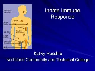
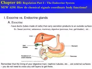
Organs of the Immune System • Bone marrow-site of hematopoiesis, B cell education, no afferent lymphatic vessels (no vessels coming in) • Thymus-site of T cell education, no afferent vessels • Spleen-immune response to blood borne pathogens • Lymph nodes-afferent and efferent vessels • Mucosal associated lymph tissue-afferent/efferent vessels (ie GALT)
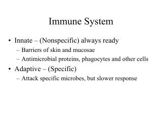
Immunity • Resistance to disease • Immune system has two intrinsic systems • Innate (nonspecific) defense system • Adaptive (specific) defense system
Immunity • Innate defense system has two lines of defense • External • First line of defense is external body membranes (skin and mucosae); provide a physical barrier of protection • Internal • Second line of defense is antimicrobial proteins, phagocytes, and other cells • Inhibit spread of invaders • Inflammation is its most important mechanism
Immunity • Adaptive defense system • Third line of defense precisely attacks foreign substances (T cells and B cells) • Takes longer to react than the innate system • Innate and adaptive defenses are deeply intertwined
Surface barriers • Skin • Mucous membranes Innate defenses Internal defenses • Phagocytes • NK cells • Inflammation • Antimicrobial proteins • Fever Humoral immunity • B cells Adaptive defenses Cellular immunity • T cells Figure 21.1
Innate Defenses • Surface barriers • Skin, mucous membranes, and their secretions • Physical barrier to most microorganisms • Keratin in skin is resistant to weak acids and bases, bacterial enzymes, and toxins • Mucosae provide similar mechanical barriers
Surface Barriers • Protective chemicals inhibit or destroy microorganisms • Skin acidity inhibits bacterial growth • Lipids in sebum and dermcidin in sweat kill bacteria • HCl and protein-digesting enzymes of stomach mucosae kill organisms • Lysozyme of saliva and lacrimal fluid • Mucus traps microorganisms
Surface Barriers • Respiratory system modifications • Mucus-coated hairs in the nose • Cilia of upper respiratory tract sweep dust- and bacteria-laden mucus from lower respiratory passages
Innate Immunity • People taking 4 or more antibiotics per year have increased risk for cancer. • Protection of cancer by isoflavones depends on ability of commensal bacteria to make them after soy ingestion
Internal Innate Defenses: Cells and Chemicals • Necessary if microorganisms invade deeper tissues • Phagocytes • Natural killer (NK) cells • Inflammatory response (macrophages, mast cells, WBCs, and inflammatory chemicals) • Antimicrobial proteins (interferons and complement proteins) • Fever
Phagocytes • Phagocytes • Macrophages • Neutrophils
Phagocytes • Macrophages develop from monocytes to become the chief phagocytic cells • Free macrophages wander through tissue spaces • E.g., alveolar macrophages in the lungs • Fixed macrophages are permanent residents of some organs • E.g., Kupffer cells (liver) and microglia (brain)
Phagocytes • Neutrophils • Become phagocytic on encountering infectious material in tissues
Mechanism of Phagocytosis Step 1: Adherence of phagocyte to pathogen • Facilitated by opsonization—coating of pathogen by complement proteins or antibodies (“BBQ sauce”, makes the pathogen tasty to immune cells)
Innate defenses Internal defenses (a) A macrophage (purple) uses its cytoplasmicextensions to pull spherical bacteria (green) toward it. Scanning electron micrograph (1750x). Figure 21.2a
1 Phagocyte adheres to pathogens or debris. 2 Phagocyte forms pseudopods that eventually engulf the particles forming a phagosome. Phagosome (phagocytic vesicle) Lysosome 3 Lysosome fuses with the phagocytic vesicle, forming a phagolysosome. Acid hydrolase enzymes 4 Lysosomal enzymes digest the particles, leaving a residual body. 5 Exocytosis of the vesicle removes indigestible and residual material. (b) Events of phagocytosis. Figure 21.2b
Mechanism of Phagocytosis • Phagocytes destroy pathogens through: • Defensins (in neutrophils), antimicrobial peptides, pierce pathogen’s membrane • Acidification and digestion by lysosomal enzymes • Some pathogens resistant to lysosomal enzymes (TB) • In this case, helper T cells release chemicals that stimulate macrophage to produce a respiratory burst
Mechanisms of Phagocytosis • Respiratory burst • Release of cell-killing free radicals (including superoxide) • Activation of additional enzymes through pH change • Oxidizing chemicals (e.g. H2O2)
Natural Killer (NK) Cells • Large granular lymphocytes • Target cells that lack “self” cell-surface receptors (e.g., cancer cells) • NK cells are non-specific cells, unlike T cells which react to specific infected cells • Induce apoptosis in target cells through direct contact (not phagocytes) • Secrete potent chemicals that enhance the inflammatory response
Inflammatory Response • Triggered whenever body tissues are injured or infected • Prevents the spread of damaging agents • Disposes of cell debris and pathogens • Sets the stage for repair
Inflammatory Response • Cardinal signs of acute inflammation: • Redness (rubor) • Heat (calor) • Swelling (tumor) • Pain (dolor) (And sometimes 5. Impairment of function)
Inflammatory Response • Inflammatory mediators • Histamine (from mast cells) • Blood proteins • Kinins, prostaglandins (PGs), leukotrienes, and complement • Released by injured tissue, phagocytes, lymphocytes, basophils, and mast cells
Vasodilation and Increased Vascular Permeability • Inflammatory chemicals cause • Dilation of arterioles (aka vasodilation), resulting in hyperemia (congestion with blood), redness and heat • Increased permeability of local capillaries and edema (leakage of exudate), swelling and pain • Releases exudate, fluid containing clotting factors and antibodies, from the vessels into the interstitial space • Causes swelling (edema) and pain (increased pressure on nerve endings)
Pain is also caused by bacterial toxins and the sensitizing effect of released prostaglandins and kinins • Aspirin and some other anti-inflammatory drugs are analgesic because they inhibit prostaglandin synthesis
Inflammatory Response: Edema • Functions of the surge of exudate • Moves foreign material (toxins, bacteria, etc.) into lymphatic vessels for processing by lymph nodes • Delivers clotting proteins to form a clot, which: • acts as a scaffold for repair • isolates the area preventing bacteria, etc. from spreading
Innate defenses Internal defenses Tissue injury Release of chemical mediators (histamine, complement, kinins, prostaglandins, etc.) Release of leukocytosis- inducing factor Leukocytosis (increased numbers of white blood cells in bloodstream) Vasodilation of arterioles Increased capillary permeability Attract neutrophils, monocytes, and lymphocytes to area (chemotaxis) Leukocytes migrate to injured area Local hyperemia (increased blood flow to area) Capillaries leak fluid (exudate formation) Margination (leukocytes cling to capillary walls) Initial stimulus Physiological response Signs of inflammation Diapedesis (leukocytes pass through capillary walls) Leaked clotting proteins form interstitial clots that wall off area to prevent injury to surrounding tissue Leaked protein-rich fluid in tissue spaces Result Phagocytosis of pathogens and dead tissue cells (by neutrophils, short-term; by macrophages, long-term) Heat Redness Pain Swelling Temporary fibrin patch forms scaffolding for repair Locally increased temperature increases metabolic rate of cells Possible temporary limitation of joint movement Pus may form Area cleared of debris Healing Figure 21.3
Phagocyte Mobilization • After inflammation starts, phagocytes are mobilized to the injured site • Steps for phagocyte mobilization • Leukocytosis: leukocytosis-inducing factors from injured cells stimulates release of neutrophils from bone marrow • Margination: neutrophils cling to the walls of capillaries in the inflamed area • Diapedesis: neutrophils flatten and squeeze out of capillaries • Chemotaxis: inflammatory chemicals (chemotactic agent) given off by injured tissue promote movement toward the injured tissue
Innatedefenses Internaldefenses Inflammatorychemicalsdiffusingfrom theinflamed siteact as chemotacticagents. Capillary wall Basementmembrane Endothelium 1 Leukocytosis.Neutrophils enter bloodfrom bone marrow. Figure 21.4, step 1
Innatedefenses Internaldefenses Inflammatorychemicalsdiffusingfrom theinflamed siteact as chemotacticagents. Capillary wall Basementmembrane Endothelium 1 2 Leukocytosis.Neutrophils enter bloodfrom bone marrow. Margination.Neutrophils clingto capillary wall. Figure 21.4, step 2
Innatedefenses Internaldefenses Inflammatorychemicalsdiffusingfrom theinflamed siteact as chemotacticagents. Capillary wall Basementmembrane Endothelium 1 2 3 Leukocytosis.Neutrophils enter bloodfrom bone marrow. Margination.Neutrophils clingto capillary wall. Diapedesis.Neutrophils flatten andsqueeze out of capillaries. Figure 21.4, step 3
Innatedefenses Internaldefenses Inflammatorychemicalsdiffusingfrom theinflamed siteact as chemotacticagents. 4 Chemotaxis.Neutrophilsfollow chemicaltrail. Capillary wall Basementmembrane Endothelium 1 2 3 Leukocytosis.Neutrophils enter bloodfrom bone marrow. Margination.Neutrophils clingto capillary wall. Diapedesis.Neutrophils flatten andsqueeze out of capillaries. Figure 21.4, step 4
Phagocyte Mobilization • Monocytes follow neutrophils • after 12 hours of being in the blood stream they become macrophages with large numbers of lysosomes • Replace neutrophils on the battlefield • Macrophages are the most important cell in getting rid of cell debris as acute inflammation subsides
Infections • Pus • Mixture of dead, or dying neutrophils, broken down tissues, and living and dead pathogens. • If inflammatory mechanisms fail to clear the area of debris, collagen fibers can be laid down, walling off the sac of pus, forming an abscess
Infections • Some bacteria, like tuberculosis bacilli, cannot be digested by the macrophages that engulf them (antibiotics do not work well because TB is inside the macrophage). • Body will wall off infected cells and form granulomas (center of diseased macrophages surrounded by fibrous capsule) • Pathogens can remain walled off for years and the patient will have no clinical sxs
Antimicrobial Proteins • Enhance our innate defenses by attacking microorganisms directly or by hindering their ability to reproduce • Most important are • Interferons • Complement
Interferons • Functions • Anti-viral • Are released by infected cells and diffuse to nearby healthy cells • Stimulate cells to synthesize proteins that “interfere” with with viral replication in healthy cells • Reduce inflammation • Activate macrophages and mobilize NK cells
Innate defenses Internal defenses Virus 1 Virusenters cell. Viral nucleic acid Nucleus Host cell 2Binds interferon from cell 1; interferon induces synthesis ofprotective proteins Host cell 1Infected by virus;makes interferon;is killed by virus Figure 21.5, step 1
Innate defenses Internal defenses Virus 1 Virusenters cell. Viral nucleic acid 2 Interferongenes switch on. DNA Nucleus Host cell 2Binds interferon from cell 1; interferon induces synthesis ofprotective proteins Host cell 1Infected by virus;makes interferon;is killed by virus Figure 21.5, step 2
Innate defenses Internal defenses Virus 1 Virusenters cell. Viral nucleic acid 2 Interferongenes switch on. DNA Nucleus mRNA 3 Cell producesinterferonmolecules. Interferon Host cell 2Binds interferon from cell 1; interferon induces synthesis ofprotective proteins Host cell 1Infected by virus;makes interferon;is killed by virus Figure 21.5, step 3
Innate defenses Internal defenses Virus 1 Virusenters cell. Viral nucleic acid 2 Interferongenes switch on. DNA Nucleus mRNA 4 Interferonbindingstimulates cell toturn on genes forantiviral proteins. 3 Cell producesinterferonmolecules. Interferon Host cell 2Binds interferon from cell 1; interferon induces synthesis ofprotective proteins Host cell 1Infected by virus;makes interferon;is killed by virus Figure 21.5, step 4
Innate defenses Internal defenses Virus 1 Virusenters cell. New viruses Viral nucleic acid 5 Antiviralproteins blockviralreproduction. 2 Interferongenes switch on. DNA Nucleus mRNA 4 Interferonbindingstimulates cell toturn on genes forantiviral proteins. 3 Cell producesinterferonmolecules. Interferon Host cell 2Binds interferon from cell 1; interferon induces synthesis ofprotective proteins Host cell 1Infected by virus;makes interferon;is killed by virus Figure 21.5, step 5