بسم الله الرحمن الرحيم
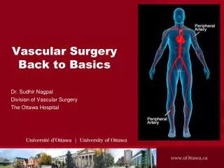
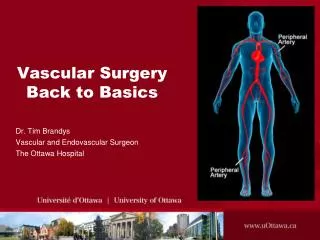
بسم الله الرحمن الرحيم. Vascular Malformations. Dr.Essam El-Kady FRCS England Head of Vascular Department Maadi Armed forces Hospital. INTRODUCTION .


بسم الله الرحمن الرحيم
E N D
Presentation Transcript
بسم الله الرحمن الرحيم DR.ESSAM EL-KADY---FRCS
VascularMalformations Dr.Essam El-Kady FRCS England Head of Vascular Department Maadi Armed forces Hospital DR.ESSAM EL-KADY---FRCS
INTRODUCTION • Vascular anomalies are among the most common congenital abnormalities observed in infants and children. Unfortunately, these lesions are also among the most confusing and misunderstood conditions, largely because of a history of inconsistent terminology used for classification. DR.ESSAM EL-KADY---FRCS
CLASSIFICATION • Hemangiomas • Vascular Malformations: • High-flow Vascular Malformation • AVMs arteriovenous malformations • AVFs arteriovenous fistulas • Low-flow Vascular Malformation • VenousMalformations • LymphaticMalformations Malformations • LymphaticVenousMalformations DR.ESSAM EL-KADY---FRCS
PATHOPHYSIOLOGY • Hemangiomas: Hemangiomas are benign tumors of infancy are grouped as • infantile hemangiomas. • rapidly involuting congenital hemangiomas. • noninvoluting hemangiomas. • intramuscular hemangiomas. • Kaposiform hemangioendothelioma (KHE). DR.ESSAM EL-KADY---FRCS
PATHOPHYSIOLOGY • Vascular Malformations: • High-flow Vascular Malformations • Arteriovenous malformations are considered to be congenital vascular anomalies, but are usually first noted several years after birth or after certain triggering changes such as trauma or the hormonal changes of puberty or pregnancy. • Arteriovenous fistulas (AVFs) are simple arteriovenous connections. Most AVFs are secondary to penetrating injuries after birth, although some are believed to be congenital. DR.ESSAM EL-KADY---FRCS
PATHOPHYSIOLOGY • Vascular Malformations: • Low-Flow Vascular Malformations • Venous malformations are spongy, masslike lesions composed of abnormal veins, ie, veins with a relative lack of smooth muscle cells in their walls DR.ESSAM EL-KADY---FRCS
PATHOPHYSIOLOGY • Vascular Malformations: • Low-Flow Vascular Malformations • Lymphatic malformations (Classified by MRI to) • Micro cystic lymphatic malformations consist of mass like soft-tissue abnormalities. • Macro cystic lymphatic malformations consist of visible cystic spaces that contain lymphatic fluid. • Mixed • Lymphatic venous malformations are a combination of abnormal lymphatic and venous channels. • Capillary malformations (port-wine stains) are common birthmarks and involve only the superficial tissues (skin) DR.ESSAM EL-KADY---FRCS
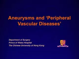
OVERGROWTH SYNDROMES: • Klippel-Trénaunay syndrome which is a low-flow combined vascular anomaly (capillary-lymphatic-venous malformation) usually associated with marked overgrowth of the leg and capillary stains. • Parkes-Weber syndrome consists of an AVM-like high-flow malformation that involves the entire extremity (usually a lower limb), and it is usually associated with a capillary malformation over the enlarged limb. DR.ESSAM EL-KADY---FRCS
Klippel-Trénaunay syndrome DR.ESSAM EL-KADY---FRCS
SIGNS AND SYMPTOMS • Hemangiomas • usually first appear a few weeks after birth and affect the head and neck in most patients. The trunk and extremities are less commonly involved. Hemangiomas look like red, flat or raised, patches or plaques with or without a cluster of superficial veins.Hemangiomas are generally firm and rubbery to the touch. DR.ESSAM EL-KADY---FRCS
Hemangiomas DR.ESSAM EL-KADY---FRCS
SIGNS AND SYMPTOMS • Vascular Malformations: • High-flow Vascular Malformations • Arteriovenous malformations (AVMs) are generally present in neonates at birth, but they often suddenly become obvious when the patient is older because of various stimuli such as trauma, pregnancy, or puberty. There are four recognized stages of AVMs: • Stage I lesion has a pinkish-bluish stain and warmth. • Stage II, the lesion has pulsations, thrill, and bruit. • Stage III, the patient has dystrophic skin changes, ulceration, bleeding, and pain. • Stage IV, the patient has high-output cardiac failure. DR.ESSAM EL-KADY---FRCS
High-flow Vascular Malformations DR.ESSAM EL-KADY---FRCS
SIGNS AND SYMPTOMS • Vascular Malformations: • Low-Flow Vascular Malformations • Venous malformations: are congenital lesions but usually become symptomatic in older children or young adults, with bluish skin discoloration, local swelling, and pain. Although venous malformations are considered benign entities, some extensive venous malformations can result in significant morbidity, particularly those in the head and neck (eg, with airway involvement). Extremity venous malformations may be associated with a limb-length discrepancy, particularly if the malformation is large. Venous malformations of the gastrointestinal tract most commonly cause chronic bleeding and anemia. DR.ESSAM EL-KADY---FRCS
Low-Flow Vascular Malformations DR.ESSAM EL-KADY---FRCS
SIGNS AND SYMPTOMS • Vascular Malformations: • Low-Flow Vascular Malformations • Lymphatic malformations commonly occur in the cervicofacial region (approximately 75% of lymphatic malformations). Most lymphatic malformations are apparent in young children. These malformations appear in various forms, such as localized small lesions or in the diffuse involvement of an extremity or particular body part or organ system. The overlying skin can be normal, or it may have tiny characteristic vesicles. Lymphatic malformations in an extremity can cause diffuse or localized swelling with soft-tissue and skeletal overgrowth DR.ESSAM EL-KADY---FRCS
Lymphatic malformations DR.ESSAM EL-KADY---FRCS
SIGNS AND SYMPTOMS • Vascular Malformations: • Low-Flow Vascular Malformations • Lymphatic venous malformations (LVMs) consist of mixed clinical and imaging findings of lymphatic malformations and venous malformations. DR.ESSAM EL-KADY---FRCS
DIAGNOSIS • Most vascular anomalies, particularly the superficial anomalies (eg, capillary malformations port-wine stains) are recognized by simple clinical history and clinical assessment and do not require any imaging studies. However, most anomalies extending into the deep tissues require imaging studies • To confirm the initial diagnosis, • To determine the extent of the malformation • To plan treatment DR.ESSAM EL-KADY---FRCS
DIAGNOSIS • MRI IS THE IMAGING STUDY OF CHOICE • Angio- MRA- CT angio: The gold standard for high-flow anomalies is conventional arteriography,however the new noninvasive angiographic techniques such as magnetic resonance angiography or computed tomographic angiography offer noninvasive assessment of the flow dynamics and vasculature of high-flow anomalies (eg, arteriovenous malformation, arteriovenous fistula). • Duplex ultrasonography: Portability and availability are the main advantages of ultrasonography compared with MRI. Ultrasonography is commonly used to quickly evaluate anomalies during the patient's initial visit to confirm the suspected diagnosis. It is also used to triage patients and schedule them for appropriate treatment. DR.ESSAM EL-KADY---FRCS
TREATMENT • Hemangiomas: Most hemangiomas regress gradually and require no treatment • Surgical Treatment: excision of the localised hemangioma • Radiotherapy/ Arterial embolization: can be used in selected cases. • Medical Treatmen • The leading pharmacologic agents used for hemangiomas are steroids, either by systemic use or intralesional injection. • Angiogenesis inhibitors such as interferon, vincristine, can be used in selected cases. DR.ESSAM EL-KADY---FRCS
Surgical Treatment of Hemangioma DR.ESSAM EL-KADY---FRCS
TREATMENT • Low-flow Malformations: • Surgical treatment :a few patients with venous malformations can be treated with a simple surgical excision especially if small and dose not involve vital structure. • Sclerotherapy: most patients with venous malformation are dependent on sclerotherapy (in which we infuse sclerosant agent into the lesion under various imaging guidance techniques). Currently, the most commonly used sclerosant agent is absolute alcohol. Other, less commonly used agents, include ethanolamine oleate (Ethamolin) and sodium tetradecyl sulfate (Sotrecol). DR.ESSAM EL-KADY---FRCS
Treatment of Low-flow Malformations: DR.ESSAM EL-KADY---FRCS
TREATMENT • High-flow Malformations: • Surgical treatment: Small, superficial arteriovenous malformations can be removed surgically. However, • Embolization: It has been the only feasible treatment option for most arteriovenous malformations. Embolization, which closes off the arterial feeders of the malformation, is generally effective in arteriovenous malformations to stabilize the malformation. DR.ESSAM EL-KADY---FRCS
OUTCOMES Although they often cause significant psychosocial stress for parents and potentially for children, most vascular anomalies are benign conditions and do not require diagnostic tests or treatments. However, some (eg, arteriovenous malformations or large venous malformations) are quite problematic, causing significant discomfort or disability, and they may worsen. Unfortunately, misclassifications or incorrect diagnoses are common and usually a result of the limited experience of the clinicians or radiologists involved in the diagnosis and management. With the appropriate diagnostic workup and therapeutic management, even rapidly progressing malformations can be managed successfully. DR.ESSAM EL-KADY---FRCS
THANK YOU DR.ESSAM EL-KADY---FRCS