Diffraction Small Features ( ) Reciprocal Space: k x = 2/x Ordered Array
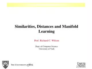
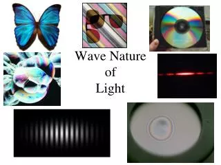
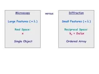
Microscopy Large Features ( » ) Real Space: x Single Object . Diffraction Small Features ( ) Reciprocal Space: k x = 2/x Ordered Array. versus. Real space versus reciprocal space. y. k y. k x = 2/x x = row spacing k x rows. Reciprocal space: k x , k y , k z.

Diffraction Small Features ( ) Reciprocal Space: k x = 2/x Ordered Array
E N D
Presentation Transcript
MicroscopyLarge Features (»)Real Space:x Single Object DiffractionSmall Features ()Reciprocal Space:kx = 2/xOrdered Array versus
Real space versus reciprocal space y ky kx = 2/xx = row spacingkx rows Reciprocal space: kx,ky,kz Real space: x,y,z
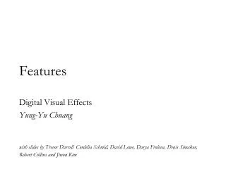
Test patterns for simulating diffraction from DNA Single helix Double helix
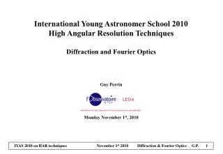
Rosalind Franklin’s x-ray diffraction pattern of DNA, which led to the double-helix model(Linus Pauling’s copy)
X-ray diffraction pattern of DNA Diffraction pattern The double helix of DNA 2 b 2 p p b p = period of one turn b = base pair spacing = slope of the helix
X-ray diffraction image of the protein myoglobin • This image contains about 3000 diffraction spots. All that information is needed to determine the positions of thousands of atoms in myoglobin. • Protein crystallography has become essential for biochemistry, because the structure of a protein determines its function.
Real space versus reciprocal space • Diffraction patterns live in reciprocal space, which corresponds to the projection screen. A direction of a beam in real space becomes a point on the screen in reciprocal space. • Everything is backwards in reciprocal space: A large distance x in real space becomes a small k-vector kx in reciprocal space and vice versa. • Even physicists have a hard time thinking in reciprocal space. But it is used widely for characterizing waves, particularly electron waves in solids and nanostructures.
Low Energy Electron Diffraction (LEED) at surfaces K= 2/d k= 2/D D d 1D chain structure 2D planar structure
a Rg P Q1/2 1/Rg Q Neutron Diffraction: Small Angle Neutron Scattering (SANS) Good for light elements (hydrogen, deuterium, polymers) and for magnetic materials (magnetic moment of the neutron). Model of a polymer: Rg = radius of gyration a persistence length (see Lecture 2 on length scales) Diffracted neutron intensity P plotted versus the k-vector Q