Download
1 / 25
260 likes | 281 Vues
This lecture covers the structural characteristics of red blood cells (RBCs), emphasizing their small size, biconcave shape, and role in transporting hemoglobin. It delves into erythropoiesis, the process of RBC production, and the destruction of erythrocytes. Key topics include the absence of a nucleus in RBCs, hemoglobin's function, and sites of RBC formation in different stages of life. The lecture also explores how erythropoiesis is crucially regulated to maintain a balance between RBC production and destruction.

E N D
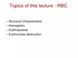
Topics of this lecture : RBC • Structural characteristics • Hemoglobin • Erythropoiesis • Erythrocytes destruction
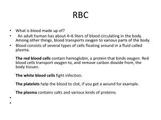
Structural characteristics • Its small size and biconcave shape provides more surface area than other spherical cells. Why is this important? • Major function of RBC is to transport hemoglobin. • Erythrocytes contain mainly hemoglobin. This is why RBC could be called “bags” of hemoglobin. Why hemoglobin has to be inside RBC and not free in plasma? 1 µm 7.8
Erythrocytes / Red blood cells • Biconcave discs, mean diameter ~7.8 µm and thickness of 2.5 / 1 µm • Typical concentration is 4.7+/- 0.3 million per cubic mm (µl) in females and 5.2+/- 0.3 million per cubic mm (µl) in males • Average volume (MCV) 90 – 95 µm3 • Lack a nucleus (average lifespan = about 120 days) • Transport hemoglobin (each RBC has about 280 million hemoglobin molecules) • Contain carbonic anhydrase (critical for transport of carbon dioxide)
Extra information about RBCs • Each of us has 25 to 30 trillion RBCs streaming through our vessels. • They are replaced at the average rate of 2 to 3 million cells per second. • Without DNA and RNA the RBCs cannot synthesize proteins for repair, growth and renewal of enzymes. • During its life each RBC travels about 700 miles as it circulates through the vasculature.
Structural Characterstics of RBC ** No nucleus (anucleate) or organelles (no mitochondria, no endoplasmic reticulum) In the RBC cytosol there are different proteins such as: Hemoglobin – made before loss of nucleus Not only carries oxygen but also acts as protein buffer Spectrin – promote changes in RBC shape Enzymes: for Forming ATP from glucose metabolism Maintaining flexibility (elasticity) of the cell membrane Transport ions across the cell membrane Keeping iron in ferrous state Acting as antioxidants Note: when these enzymes become less active in old RBCs, the cells become more fragile and RBCs rupture during their passing through tight capillaries (specially in spleen). ***Splenctomy leads to increased no. of abnormal shaped RBCs in circulation.
Where are Erythrocytes produced? • In early weeks of pregnancy, a primitive nucleated RBC are formed in yolk sac • Middle trimester of fetal life- Liver (mainly), spleen, lymph nodes. • Last month of pregnancy and after birth- exclusively from Bone marrow Sites of RBC formation in different ages 0-5 Y …..all bones of the body 5-20 Y…. The shaft of long bones become fatty and its contribution to form RBC reduced gradually and stops completely after 20 y. Heads of long bones continue to form RBC After 20 Y….. Almost in membranous bones Relative rates of RBC production in bone marrow of different bones at different ages
Hemoglobin • It’s the protein that makes RBC red. • Binds easily and reversibly to oxygen • Oxygen moves in the blood bound to hemoglobin • Average normal values of hemoglobin • 16 g/100ml of blood in adult males • 14 g/100ml of blood in adult females
Erythropoiesis means erythrocytes production Although the various formed elements have different functions, they all arise from the same stem cell. Pluripotent hematopoietic stem cell (PHSC) Derived from mesenchyme – once committed it follows path PHSC gives to 1.Committed stem cells that produce RBC, granulocytes, monocytes, plataelts 2. Committed stem cells that give lymphocytes 3. PHSC that keep supply of committed stem cells *** note: growth inducers like interleukin-3 makes PHSC to give different committed stem cells
***Note: differentiationof different colony into different blood cells is done by different inducers Committed stem cell that form RBC, Granulocytes, monocytes, platelets Committed stem cell that form lymphocytes
Erythropoiesis* Ertyhropoiesis begins when a stem cell is transformed into a proerythroblast. Proerythroblast is the first cell belonging to red blood series. At early stages of erythroblast, little of Hb starts to accumulate and in the late stages of erythroblast formation Hb concentration is increased until it forms almost 34% of volume of the cell. Very few Hb is formed by reticulocytes.
Stages of RBC Maturation* • Committed stem cell • Proerythroblast • Erythroblast • Reticulocyte • Mature RBC The last stage of development is called reticulocytes which do not contain nucleus and the cytoplasmic organelles are disappearing and only remnants of these remained. Reticulocytes leave bone marrow and stay in blood for 1-2 days and finally they form mature RBC. Normal reticulocytes no. in circulation is about 1% of total RBC no. BONE MARROW Time needed for committed stem cellsto develop to mature erythrocytes is about 5-7 days.
Erythropoiesis Erythropoiesis needs to be controlled so there is a balance between RBC production and destruction. New cells are made at a rate of more than 2 million per second in healthy people. This process is controlled hormonally and depends on adequate supplies of iron, amino acids,Vit. B12 and folic acid.
Erythropoiesis Control Erythropoietin – glycoprotein with MW of 34000. There is always a small amount of this hormone in the blood keeping a basal rate of production of RBC Produced mainly by the kidneys (90%) but the liver produces some (10%). A drop in normal oxygen levels (hypoxia) triggers erythropoietin formation Hypoxia is most potent stimulus for erythropoietin production. Another factors increases erythropoietin production: Androgen alkalosis Catacholamines Too many erythrocytes depresses erythropoietin production.
IMBALANCE Homeostasis: Normal blood oxygen levels 1 Stimulus: Hypoxia (low blood O2- carrying ability) due to • DecreasedRBC count • Decreased amountof hemoglobin • Decreasedavailability of O2 5 O2- carryingability of bloodincreases. IMBALANCE 4 Enhancederythropoiesisincreases RBCcount. 2 Kidney (and liver toa smaller extent)releaseserythropoietin. 3 Erythropoietinstimulates redbone marrow. Figure 17.6, step 5
Erythropoietin production and effects ***Produced by the tubular epithelial cells in kidney and hepatocytes in the liver ***EFFECTS • Increases number of proerythroblasts • Stimulates red bone marrow to increase rate of cells division.
Destruction of Erythrocytes The anucleate condition of erythrocytes carries with it some important limitations. Red blood cells are unable to synthesize new proteins, to grow, or to divide. Erythrocytes become “old” as they lose their flexibility and become increasingly rigid and fragile, and their contained hemoglobin begins to degenerate. Aged RBCs have: Metabolic activity Enzyme activity ATP Membrane Lipids Fragile Membrane Cells Rupture as they pass through narrow spaces in spleen
Destruction of Erythrocytes Red blood cells have a useful life span of 100 to 120 days, after which they become trapped and fragment in smaller circulatory channels, particularly in those of the spleen. In the red pulp of the spleen, RBC rupture when they try to squeeze through because of their fragile old membrane. For this reason, the spleen is sometimes called the “red blood cell graveyard”.
Erythrocyte Destruction Macrophages in spleen, liver and red bone marrow phagocytize dying RBC. Globin – breaks into amino acids, which can be reused to produce other proteins Heme – iron and porphyrin Fe – removed and recycled in spleen Porphyrin – converted to bilirubin (bile pigment) Yellow pigment secreted by liver into bile, which is excreted in urine and feces
Anemia • Any decrease in blood’s oxygen-carrying capacity is known as anemia. • Causes: • Insufficient number of RBC • Low hemoglobin content • Abnormal hemoglobin • One of the major effects of anemia is the greatly increased work load on the heart.
1-Blood loss : Microcytic , hypochromic an. No enough iron to form Hb 2-Aplastic An BM DIS3-Megaloplastic, Large RBCDec Vit B12,folic acidand Intrinsic factor4-Pernicious anemia caused by atrophy of stomach mucosa or gastrectomy can lead to megaloplastic anemia ANEMIA
4-Hemolytic anemia a-Hereditary, Spherocytosis b-Sickle cell (HbS HB) c-Erythroblastosis fetalis Rh+ fetus with AB from mother Rh-d-Thalassaemia :Inherited impairment of Hb production (Minor & major)
Effect of anemia on cardiovascular system** Decreased viscosity anemia Decreased resistance to blood flow More blood returns to the heart hypoxia Dilatation of blood vessels More cardiac output
Polycythemia** Primary polycythemia (polycythemia vera) Due to increased activity of hemocytoblastic cell of bone marrow polycythemia (Means increased RBCs no.( Secondary polycythemia Due to hypoxia
Effects of polycythemia on CVS* Increased cardiac output Polycythemia Leads to Increased venous return Blood volume Hematocrit viscosity decreased blood flow Increased blood pressure Decreased venous return to the heart More O2 is extracted from Hb and thus deoxygenated blood is increased leading to bluish discoloration of the skin (cyanosis)) Decreased cardiac output