Download

1 / 47
480 likes | 948 Vues
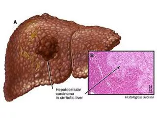
Definition. hepatocellular carcinoma (HCC)-- derived from hepatocytes cholangiocarcinoma (CC)-- arising from intrahepatic bile duct epitheliumcombined hepatocellular and cholangiocarcinoma (cHCC-CC)-- involving both hepatocellular and cholangiocellular components in the same tumor .

E N D
1. Primary Liver Carcinoma Chen Yingxuan
Department of Gastroenterology,Renji Hospital
Shanghai Institute of Digestive Diseases
2. Definition hepatocellular carcinoma (HCC)--
derived from hepatocytes
cholangiocarcinoma (CC)--
arising from intrahepatic bile duct epithelium
combined hepatocellular and cholangiocarcinoma (cHCC-CC)--
involving both hepatocellular and cholangiocellular components in the same tumor
3. Morphological classification lump lesions
nodular lesions
Diffused lesions
4. Distribution of HCC
5. Incidence(per 100,000 population) of HCC in China
6. Aetiology Hepatitis B virus
Hepatitis C virus
Liver cirrhosis
Aflatoxins (AF)
Others
7. Clinical Presentation
8. Symptoms-early stage No symptom/No specific symptoms
Pain
Poor appetite
Early satiety
Abdominal swelling
Weight loss
Weakness, tiredness
An awareness of a lump in the upper abdomen
Fever
9. Presentation with symptoms of advancing cirrhosis
Pruritus
Jaundice
Variceal bleeding
Cachexia
Hepatic encephalopathy
Increasing abdominal girth (portal vein occlusion by thrombus or tumor associated with rapid onset of ascites)
10. Physical Findings Hepatomegaly
Jaundice
Ascites
Splenomegaly
spider angiomata
Pedal edema
Periumbilical collateral veins
Enlarged hemorrhoidal veins
11. hypoglycemia
polycythemia
hypercalcemia Paraneoplastic Manifestations
12. Complications Hepatic encephalopathy
Gastrointestinal bleeding
Cancer rupturing and bleeding into the abdominal cavity
Infection
13. Diagnosis
14. Serum Tumor markers Alpha-fetoprotein (AFP)
an a1-globulin normally present in high concentration in fetal serum but in only minute amounts thereafter
Normal levels < 10 ng/ml
Elevation:75% of HCC cases, active liver disease, embryonal cell carcinomas, metastatic cancer in the liver
15. Alpha-fetoprotein (AFP)-clinical significance
AFP >400?g/L, up to 1 month or AFP>200 ?g/L , up to 2 months; and excluding patients with pregnant, active liver disease, embryonal cell carcinomas
screening or primary diagnosis ( detection of HCC at an earlier stage, often before the development of symptoms)
following the response to treatment Serum Tumor markers
16. Serum Tumor markers Fucosylated a-Fetoprotein
Des-?-carboxy Prothrombin
a-l-Fucosidase
?-GT-II
18. Imaging Studies-Ultrasonography
19. CT of large HCC
20. Inoperable extensive liver metastases
21. Imaging Studies-MRI
22. Imaging Studies- Angiography
23. Biopsy This biopsy may be obtained by a fine needle under local anesthesia, using ultrasound or CT scan to guide the needle into the tumor
Lesions that are 2-3 cm or smaller may be dysplastic nodules in a cirrhotic background. These probably are premalignant, and obtaining a biopsy is especially important to distinguish them from HCC.
Obtaining a biopsy may be unnecessary in patients who will undergo resection regardless of diagnosis, such as those without cirrhosis or evidence of metastatic disease.
24. Histologic Findings
26. Clinical diagnostic standard AFP >400?g/L, excluding patients with pregnant, active liver disease, embryonal cell carcinomas and hepatic metastases, accompanied by palpable hepatic mass or imagining scan of the liver to detect for the presence of tumor nodule(s)
AFP <400?g/L, excluding patients with pregnant, active liver disease, embryonal cell carcinomas and hepatic metastases, as well as confirming the presence of the mass by using at least two imaging modalities or showing two positive serum markers and one imaging modality
The presence of the clinical presentation and signs of distant spread, excluding hepatic metastases
2001 the 8th National Liver Cancer Conference
27. Differentials Cirrhosis
Metastatic disease
Benign tumor of the liver
Liver abscess
28. Staging-TMN(Tumor, node, and metastases ) T1
Solitary tumor smaller than or equal to 2 cm
No vascular invasion
T2
Solitary tumor smaller than or equal to 2 cm, with vascular invasion
Multiple tumors, in 1 lobe only, smaller than or equal to 2 cm, no vascular invasion
Solitary tumor larger than 2 cm, no vascular invasion
T3
Solitary tumor larger than 2 cm, with vascular invasion
Multiple tumors, in 1 lobe only, with vascular invasion
T4
Multiple tumors involving more than 1 lobe
Involvement of a major branch of the portal or hepatic vein
29. Staging-TMN(Tumor, node, and metastases ) N0 - Indicates no nodal involvement
N1 - Indicates regional nodal involvement
M0 - Indicates no distant metastasis
M1 - Indicates metastasis presence beyond the liver
30. Stage grouping
Stage I = T1 + N0 + M0
Stage II = T2 + N0 + M0
Stage IIIA = T3 + N0 + M0
Stage IIIB = T1-3 + N1 + M0
Stage IVA = T4 + N0 + M0
Stage IVB = T1-4 + N0-1 + M1
31. Treatment
32. Treatment options Surgical resection and liver transplantation
Transcatheter arterial chemoembolisation (TACE)
Percutaneous Ethanol Injection
Radiofrequency ablation
Systemic treatment with chemotherapy
Radiotherapy
Others
33. Surgical The only proven potentially curative therapy for HCC
hepatic resection
liver transplantation
The main prognostic factors for resectability
tumor size and liver function
Patients with single small HCC (<=5 cm) or up to three lesions (<=3 cm )
34. hepatic resection Tumor confined to one lobe of the liver and be favorably located
The nontumorous liver tissue should not be cirrhotic
Higher success rates in tumors smaller than 2 cm with no vascular invasion (T1 N0 M0, stage I)
Resection is feasible in only about 15% of patients
A 5-year survival rate : 40%
35. Liver transplantation should be considered in any patient with cirrhosis and a small (5 cm or less single nodule or up to three lesions of 3 cm or less) HCC
the prognosis for long-term survival is poor (20-30%)
limited availability of organs and long wait times
36. producing tumor necrosis
Indications
palliation of inoperable primary or secondary hepatic malignancies and reduction of pain
Preoperation: to reduce tumor mass
Postoperation:to influence tumor rests or recurrences
Contraindications
more than 75% of the liver is involved by tumor
presence of insufficiency of the liver, significant portal hypertension, occlusion of the portal vein or hepatorenal syndrome
TACE
37. Antineoplastic scheme FAP: 5-FU 750~1000 mg
MMC 10~20 mg
CDDP 60~80 mg
FMP: 5-FU 750~1000 mg
ADR 20~40 mg
CDDP 60~80 mg
38. Before TACE
39. First TACE: HCC prior being submitted to transcatheter arterial chemoembolization. The angiographic study allows clear visualization of the hypervascular tumor
40. After second TACE: which is further studied by means of superselective catheterization
42. PEI has been shown to produce necrosis of small HCC. It is best suited to peripheral lesions, less than 3 cm in diameter
Percutaneous Ethanol Injection
43. Radiofrequency Ablation(RA) using a probe placed into the tumor mass
A single probe can destroy lesions of up to 3 cm and a multiple tipped probe has been used to target lesions of up to 6 cm in diameter
44. Systemic chemotherapy a poor response - caused by the universal expression of the multidrug resistance gene protein on the surface of the malignant cells
The most active drugs - doxorubicin, cisplatin, and fluorouracil
Response rates - under 10%
Combination chemotherapy does not add any benefit to single-agent chemotherapy
45. Radiatherapy
46. Other Treatment Methods High Intensity Focused Ultrasound (HIFU)
Chinese medicine
47. Prognosis Overall prognosis for survival depends on the extent of cirrhosis and tumor stage
curative resection - a median survival of 4 years
not to be treated - a median survival of 3 months