Download
1 / 64
670 likes | 990 Vues
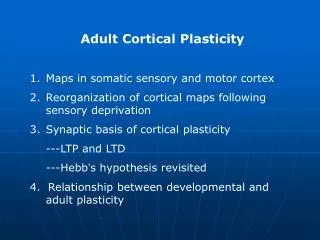
Cortical and Subcortical Motor Systems and Disorders. Nancy L. Sicotte, M.D. Director, Multiple Sclerosis Program Cedars-Sinai Medical Center Associate Professor in Residence Department of Neurology David Geffen School of Medicine at UCLA. Overview. Anatomy - Cortical

E N D
Cortical and Subcortical Motor Systems and Disorders Nancy L. Sicotte, M.D. Director, Multiple Sclerosis Program Cedars-Sinai Medical Center Associate Professor in Residence Department of Neurology David Geffen School of Medicine at UCLA
Overview • Anatomy - Cortical • Upper versus Lower Motor Neuron Changes • Anatomy - Subcortical • Syndromes • Structural/Functional Imaging Approaches
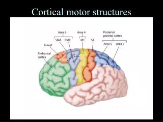
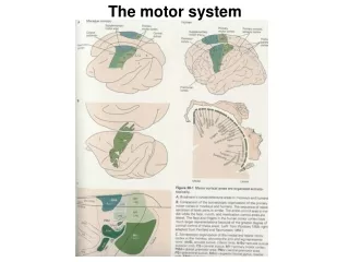
Major Cortical Motor Pathways
Motor and Sensory Tracts Through the Brainstem
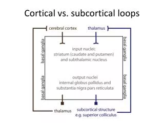
Cortical/Subcortical Connections For Motor control
Upper Motor Neuron (UMN) vs. Lower Motor Neuron (LMN) Signs * Some atrophy may develop due to disuse
Connectivity diagram showing excitatory glutamatergic pathways as red, inhibitory GABAergic pathways as blue, and modulatory dopaminergic as magenta.
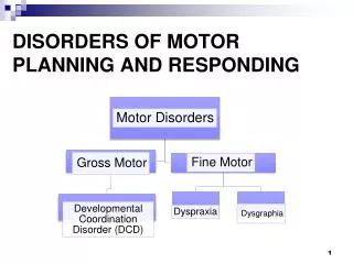
Motor Disorders • Amyotrophic Lateral Sclerosis • Multiple Sclerosis • Parkinson Disease • “Parkinson Plus” - PSP, CBD, Shy-Drager • Huntington Chorea • Tardive Dyskinesia • Spinal Cerebellar Atrophy (SCAs) • Benign Essential Tremor
ALS - “Lou Gehrig’s Disease” • “Pure” Motor System Neurodegenerative disorder • Affects Upper and Lower Motor Neurons • Onset in late middle age, Men>Women • Familial form (SOD mutation) accounts for 10% of cases • Median survival = 5 years (3 years for bulbar forms) • Cognitive impairment now recognized to occur • Pathological laughing/crying
Multiple Sclerosis • Autoimmune demyelinating disease of the central nervous system • Age of onset 20-40, affects women: men - 2:1 • Wide spread white matter lesions • Gray matter atrophy • Relapsing-remitting course usually followed by progressive myelopathy • Cognitive impairment/disinhibition common (la belle indifference)
A B A C B MS Lesions: Axonal Changes Courtesy of Rudick R
Parkinson Disease • Neurodenerative disease resulting in loss of pigmented neurons in the substantia nigra – mitochondria? • Occurs in elderly men>women • Environmental factors likely important (pesticides, MPTP) • Difficulties with initiation of movement • Tremor at rest • Depression/cognitive impairment common • Treatment: Dopamine agonists • Deep brain stimulators/stereotaxic surgery
Parkinson “Plus” • Share phenotype of tremor, stiffness, impaired initiation but with other features - many with Lewy bodies • Progressive supranuclear palsy (PSP) - eye movement impairment, axial rigidity, personality changes • Corticobasal degeneration (CBD) - present with unilateral (usually upper limb) apraxia, progressive motor/cognitive impairment, cortical lewy bodies • Dementia with Lewy bodies (DLB)- may look like Alzheimer’s, frequent hallucinations, sleep disturbance • Shy-Drager - like Parkinson disease, but severe autonomic involvement early • Drug induced Parkinsonism - very common
Huntington Chorea • Autosomal dominant disorder, first CAG repeat disorder described • Symptoms begin in 40-50s. • May present with psychiatric symptoms, depression, psychosis, mania • Caudate atrophy • Choreathetoid movements develop later • Progressive decline in function over 10-15 years • No effective treatment
Tardive Dyskinesia • Develops after long term use of neuroleptics (dopamine antagonists) • Frequently seen in patients with schizophrenia • More likely with “high potency” forms ie Haldol • Symptoms improve with use of offending agent - vicious cycle
Dystonia • Abnormal, sustained contraction of muscles - (agonists and antagonists) • Common forms - cervical, hand (writers cramp), can be generalized • Cervical dystonia (torticollis) - “sensory tricks” sometimes work • Some rare forms are responsive to dopamine agonists • Cause not well understood ?traumatic, overuse? • Can be caused acutely by certain drugs • Most common treatment now is Botox
Spinocerebellar Atrophy • Multiple genetic forms now recognized • Many with triple nucleotide repeats • Variety of phenotypes, many involving cognitive impairment • Severe Cerebellar Atrophy • Currently no effective treatments
Water Diffusion - Isotropic and Anisotropic Beaulieu, C. NMR Biomed 2002;15:435-455
Diffusion along axons is anisotropic Beaulieu, C. NMR Biomed 2002;15:435-455
Diffusion gradients applied in 6 directions Masutani et al. Eur J Radiology, 46(2003):53-66
Tensor Displayed as an Ellipsoid Masutani et al. Eur J Radiology, 46(2003):53-66
Diffusion Tensor Imaging ofMajor White Matter Tracts Pajevic and Pierpaoli, Mag Res Med (1999);42:526-540
Diffusion Tensor Imaging FIBER ORIENTATION: Red = right/left Blue = dorsal/caudal Green = anterior/posterior Myelin Stain - Mid pons
Horizontal Gaze Palsy with Progressive Scoliosis (HGGPS) Crisfield 1974 Dretakis & Kondoyannis 1974 Sharpe et al., 1975
MRI HGPPS Patient Sicotte et al Neurology, (67):519-521, 2006
Midpons - DTI Control HGPPS
Sagittal View - DTI Control HGPPS Sicotte et al Neurology, (67):519-521, 2006
Coronal View - DTI Control HGPPS Sicotte et al Neurology, (67):519-521, 2006
Tract tracing with Diffusion Tensor Imaging (DTI)
Diffusion Tensor Fiber Tracking:Corpus Callosum Huang et al. Neuroimage 26(2005):195-205
Diffusion Tensor Imaging Metrics • Mean Diffusivity (MD) - average of diffusion in all directions • Axial Diffusion (AD) - 1first eigenvalue - parallel to tract • Radial Diffusion (RD) - 2,3 average of 2nd and 3rd eigenvalues - perpendicular to tract - • Fractional Anisotropy (fA) - dimensionless number ranging from 0 (isotropic) to 1 corresponding to the degree of preferential flow
Group Differences: Fractional Anisotropy in RRMS vs Controls
fA group/correlation and with composite motor score Controls vs. RRMS Correlation with motor function