Download
1 / 15
190 likes | 468 Vues
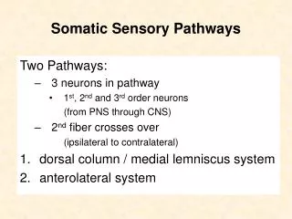
Adult Cortical Plasticity Maps in somatic sensory and motor cortex Reorganization of cortical maps following sensory deprivation Synaptic basis of cortical plasticity ---LTP and LTD ---Hebb ’ s hypothesis revisited 4. Relationship between developmental and adult plasticity.

E N D
Adult Cortical Plasticity • Maps in somatic sensory and motor cortex • Reorganization of cortical maps following sensory deprivation • Synaptic basis of cortical plasticity • ---LTP and LTD • ---Hebb’s hypothesis revisited • 4. Relationship between developmental and adult plasticity
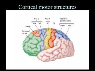
Properties of Cortical Maps • Topographically ordered: Nearby points in periphery are represented by nearby cortical neurons. • Multiple Representations: The same set of sensory or motor information are represented repeately by multiple cortical areas. • Distorted mapping: Periphery points that required higher spatial resolution are represented with disproportional cortial areas (larger number of cortical neurons).
Plasticity of rat somatosensory cortex Barrel Cortex – receiving sensory inputs from whiskersDepriving sensory inputs by removing whisker – shrinkage of corresponding barrels-- Importance of normal sensory inputs even in adult-- Activity-dependent competition exists in adult cortex Barrel cortex
Functional changes in V1 due to scotoma (blind spot) Visual field is represented by the grid on the retina, with corresponding maps shown on V1. Lesion of retina first silenced the corresponding cortical area, but reorganization of the receptive fields of cortical neurons leads to increased representation of the areas around the lesion and reduced representation of the lesioned area. (Gilbert and Wiesel) Artificial scotoma – Deprivation of visual input to specific region of retina without lesion results in similar reorganization of the cortical receptive fields.
Functional expansion of cortical representation by repetitive use Monkey was trained in a task that required heavy usage of digits 2,3,4 --expansion of cortical representation of these digits after a few months
Functional changes in the somatic sensory cortex of an owl monkey following amputation of a digit. Question remains to be answered: Are functional changes due to structural changes in the connectivity between neurons, or simply silencing of synaptic transmission, e.g., long-term depression or increased inhibition?
Evidence from human studies Functional brain imaging studies showing larger cortical representation of left figures for string player who has an earlier inception of practice, although string players in general have higher representation than non-string players (controls) in the same orchestra.
Use-dependent changes in synaptic functionsShort-term plasticity: synaptic facilitation, synaptic fatigue, post-tetanic potentiation (PTP) Facilitation (10s msec): Increased transmitter release due to residue Ca2+ of previous stimuli Fatigue (100s msec): Depletion of synaptic vesicle supply due to high-frequency use Post-tetanic potentiation (minutes): Increase mobilization of vesicle supply due to Ca2+ accumulation induced by tetanus (Found to different degrees at all synapses)
Use-dependent changes in synaptic functionsLong-term potentiation (LTP) and Long-term depression (LTD) • -- Persistent increase or decrease in synaptic response due to repetitive activity, found in hippocampus and cortex • -- Brief high-frequency stimulation – LTP • Prolonged low-frequency stimulation – LTD • Mechanism: • Induction of either LTP or LTD requires postsynaptic Ca2+ rise. • At most synapses, activation of NMDA receptors is required for • the induction of LTP/LTD. • 3. LTP/LTD at many synapses are due to increase/decrease of postsynaptic AMPA-type glutamate receptors, but presynaptic increase/decrease of transmitter release may also occur.
Developmental vs. adult plasticity • Are these two forms of plasticity depend on similar synaptic mechanisms? • Evidence: • -- Development of ocular dominance columns is prevented by blocking NMDA receptors. (M. Constantine-Paton) • -- Critical period plasticity (ocular dominance modification due to monocular deprivation) can be revived in adult primary visual cortex by protease treatment (that remove extracelluar matrix around neurons). (L. Mafei) • -- LTP/LTD can be induced in developing and adult cortex by similar stimulation. • -- LTP/LTD induction can result in structural changes at synapses, presumably also changes in connectivity • LTP – increase spine formation, swelling of existing spines • LTD – shrinkage and retraction of spines
Do learning and memory in adult brain involves processes similar to activity-dependent developmental refinement of connections? • Evidence: • -- LTP is required for spatial learning (hippocampus) and fearing conditioning (amygdala) in rats • -- LTP/LTD induction is accompanied by structural changes at synapses • -- Neruotrophins required for developmental refinement of connections (e.g., in ocular dominance segregation) is also required for LTP induction in adult brain. Neurotrophins
The key question: Does activity-induced LTP and LTD leads to formation and elimination of synaptic connection? (Does functional plasticity lead to structural plasticity? )