Motor cortex
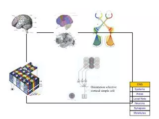
Motor cortex. Organization of motor cortex Motor cortical map Effect of cortical motor neuron activation on muscle contraction Population coding. Cortical areas involved in motor control. Cortical areas involved in motor control. (Association cortex). Primary motor cortex (M1)

Motor cortex
E N D
Presentation Transcript
Motor cortex • Organization of motor cortex • Motor cortical map • Effect of cortical motor neuron activation on muscle contraction • Population coding
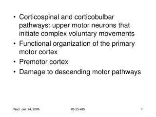
Cortical areas involved in motor control (Association cortex) Primary motor cortex (M1) - initiation and execution of movement Premotor and Supplementary motor cortex - initiation of complex movement, planning the movement Activity detected in the motor area (by fMRI) Flexing the finger -- M1 only Writing a letter with finger (complex sequence of movement) – M1, premotor and supplementary cortex Think about writing with the finger - premotor and supplementary cortex, not M1.,
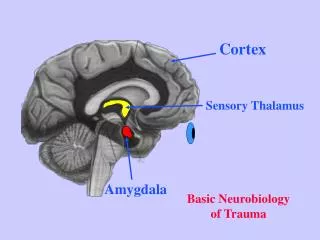
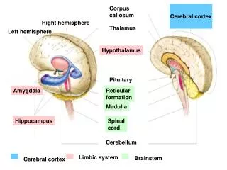
Planning, initiation of voluntary movement Sensory-motor integration, motor learning Basic movement, posture Reflex (involuntary movement)
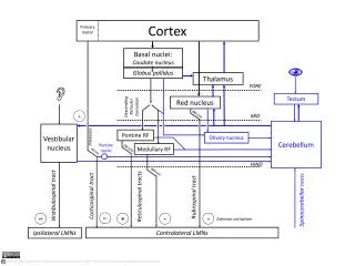
Connections between different motor areas (integrating all sensory informatio (premotor and supplementary motorareas) Cortico- spinal tract
Stimulation of motor cortex can cause muscle activity EMG (electromyogram) – recording of muscle contraction activity using extracellular or surface electrode.
Evidence for motor cortical map Intracortical stimulation • Brain stimulation • Intracortical stimulation • TMS (transcranial magnetic stimulation) • Jacksonian march (propagation of seizure activity) • - progressive activation of motor cortex • Functional Brain Imaging (detection of the active brain areas) • Positron Emission Tomography (PET) • -- Detection of activity-related glucose or O2 use by radiation due to positron emission from radioactive non-metabolizable glucose (16O, 18F labeled) or radioactive O2 (16O) • 2. Functional Magnetic Resonance Imaging (fMRI) • -- Magnetic resonance resonance of the ratio of oxygenated-nonoxygenated hemoglobin as an indication of increase flow of oxygenated blood flow to the active brain regions. TMS
Somatotopic map in primary motor cortex Distorted map: disproportionally large representation of parts requiring greater precision Somatotopic maps also exist in premotor cortex & supplementary motor cortex. Stimulation induces complex movements involving multiple joints and even bilateral movement
Divergence and convergence of cortical control of muscles: -- The same muscle is controlled by several cortical sites -- One corticospinal axon control many muscles (combinatorial control) Effectiveness of cortical stimulation at different sites • Experiment: • Microelectrode stimulation over a grid area of motor cortex • Recording from a shoulder muscle (deltoid) and a wrist muscle (ECR) • Finding: • Same muscle can be activated from multiple stimulation sites • Overlap between shoulder and wrist muscle representations • Implication: • Such overlap may allow coordination of multiple muscles for motor tasks
Use-dependent plasticity of the motor map (a) Deprivation causes reduction of representation Human hand injury Rat whisker denervation
Use-dependent plasticity of the motor map (b) Practice causes expansion of representation -- Finger opposition training – touching thumb with finger in a particular sequence. Following 3 weeks of training, fMRI showed larger cortical area activated by performing the trained sequence. -- fMRI studies showed larger cortical representation of left figures for string player who has an earlier inception of practice, although string players in general have higher representation than non-string players (controls) in the same orchestra.
Information coding by motor cortical neurons • In primary motor cortex, neuron fires before movement • Four types of neurons: • Dynamic neuron – code the rate of force • Static neuron – code steady level of force • Mixed neuron – code both rate and level of force • Directional neuron – code for direction of movement Edward V. Evarts (NIH) developed technique to record from motor cortical neuron from awake monkey performing motor tasks
Motor neuron spiking: coding force or position?Experiment: fix position of movement (wrist rotation), change force applied to the rod Wrist Extensor load Conclusion: Firing of motor cortical neurons codes the force generated by the muscle. This particular neuron recorded activates flexor muscle
“Spike triggered average” demonstrate that a single spike from a single motor neuron can exert significant effect on muscle activity Response correlated with each spike
Population coding of movement direction -Direction of the movement coded by a population of neurons, rather than a single neuron Experimental setup Georgopoulos et al., 1982
Population coding of movement direction • Actual direction of movement can be predicted by the vector sum of multiple neurons: • Each vector represents one neuron • Vector direction: preferred direction of the neuron • Vector length: firing rate of that neuron during the trial Direction tuning of individual neuron Motor cortical neurons signal both force and direction!