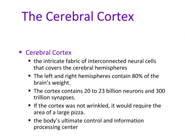
Cerebral Cortex
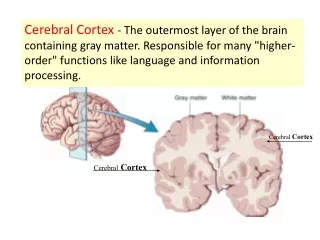
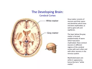
Cerebral Cortex. Cerebral Cortex. The layer of gray matter covering the entire surface of cerebral hemisphere Migration of neurons from inner mantle layer of neural tube Accommodates enormous number of neurons - Large surface area accommodates

Cerebral Cortex
E N D
Presentation Transcript
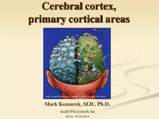
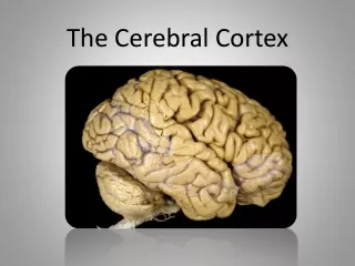
Cerebral Cortex
Cerebral Cortex • The layer of gray matter covering • the entire surface of cerebral hemisphere • Migration of neurons from inner mantle layer of • neural tube • Accommodates enormous number of neurons • - Large surface area accommodates • more neurons than deep nuclei • - Gyri and sulci also increase surface area • - Laminar organization also accommodates • enormous number of neurons
CerebralCortex Numerical Data Total surface area: 2200 cm2 (2.5 ft2) about 1/3 ------ surface area about 2/3 ------ hidden in the sulci Thickness: 1.5 mm (V I) - 4.5 mm (M I) Generally, thickest over the crest of the convolution and, thinnest in the depth of sulci Weight: 600 gm (40 % of total brain weight) 180 gm --------- neurons 420 gm --------- glial cells
CerebralCortex Numerical Data Number of neuronal cells in cerebral cortex neurons ----------- 10-15 billion glial cells ---------- 50 billion Estimation of number of cortical neurons von Economo and Koskinas (1925) 14.0 billion Shariff (1953) 6.9 billion Sholl (1956) 5.0 billion Pakkenberg (1966) 2.6 billion
Subdivision of Cerebral Cortex Allocortex Archicortex (Archipallium) Palaeocortex (Paleopallium) Isocortex Neocortex (Neopallium) cf. mesocortex, juxtallocortex, mesallocortex
Isocortex–typical 6 layered cortex I. Molecular Layer II. External Granular Layer III. External Pyramidal Layer IV. Internal Granular Layer V. Internal Pyramidal Layer VI. Polymorphic Layer
CerebralCortex Histological Organization Cellular Elements 1. Pyramidal Cell - output neuron giant pyramidal cell of Betz 2. Fusiform Cell --- modified pyramidal cell 3. Granular (Stellate) Cell basket cell, double bouquet cell, bipolar cell, chandlier cell, neurogliform cell 4. Horizontal Cell of Cajal (Retzius-Cajal cell) 5. Cells of Martinotti
CerebralCortex 1. Pyramidal Cell 2. Fusiform Cell 3. Granular (Stellate) Cell 4. basket cell 5. double bouquet cell 6. chandlier cell 7. neurogliform cell 8. Horizontal Cell of Cajal 9. Cells of Martinotti a: axon
I. Molecular Layer II. External Granular Layer III. External Pyramidal Layer Line of Kaes-Bechterew IV. Internal Granular Layer Outer band of Baillarger - Line of Gennari in area 17 V. Internal Pyramidal Layer Giant pyramidal cell of Betz Inner Band of Baillarger VI. Polymorphic Layer Golgi NisslWeigert
Cortical Afferent Fiber 1. corticocortical fiber association fiber commissural fiber 2. thalamocortical fiber- specific and non-specific 3. extrathalamic subcortical fiber cholinergic fiber - acetylcholine basal nucleus of Meynert mesolimbic dopaminergic fiber - dopamine ventral tegmental area serotonergic fiber – serotonine - raphe nuclei norepinephrinergic fiber - norepinephrine nucleus locus ceruleus
Cortical Afferent Fiber 1. association fiber 2. commissural fiber 3. specific thalamocortical fiber 4. non-specific thalamocortical fiber
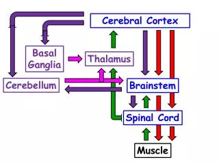
Cortical Efferent Fiber 1. Corticofugal Fiber - Projection Fiber corticostriate fiber corticothalamic fiber corticorubral fiber corticotectal fiber corticopontine fiber cortico-olivary fiber corticobulbar fiber corticospinal fiber 2. Corticocortical Fiber Association fiber Commissural fiber
Cortical Efferent Fiber 5. association fiber 6. commissural fiber 7. corticostriate fiber 8. corticorubral fiber corticopontine fiber corticobulbar fiber 9. corticospinal fiber corticotectal fiber 10. corticothalamic fiber
Columnar Cortical Unit and Cortical Circuitary A. pyramidal neuron B. excitatory granular cell C.inhibitory granular cell 1. afferent fiber 2.efferent fiber 3.corticothalamic fiber
Regional Variation of Cortical Lamination A. Homotypical isocortex ------- association cortex B. Heterotypical isocortex 1. granular cortex --- primary sensory cortex V I (17), S I (3), A I (41) 2. agranular cortex --- motor cortex M I (4), PM (6)
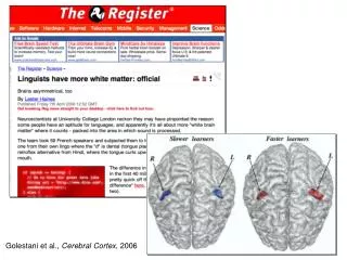
Functional Localization of Cerebral Cortex Phrenology of Gall and Spurzheim Clinical evidences Broca’s area (1861) Jacksonian epilepsy (1864) Experimental evidences Fritsch and Hitzig (1870) --- motor cortex von Gudden (1870) ---- visual cortex Ferrier (1873) ---- auditory cortex
Morphological Classification of Cortical Areas based on cytoarchitectonic studies Campbell (1905) -------- about 20 areas Brodmann (1909) ------ 47 areas - most popular Vogt and Vogt (1919) - over 200 areas von Economo (1929) -- 109 areas
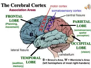
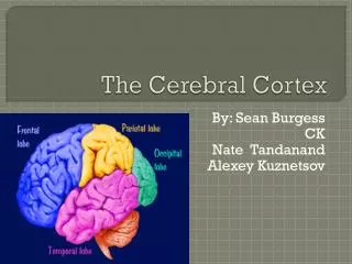
Functional Localization of Cerebral Cortex Sensory area primary sensory area secondary sensory area Motor area primary motor area secondary motor area supplementary motor area Association area parietal, occipital and temporal cortex - conceptual elaboration of sensory data prefrontal (frontal) cortex - judgement, foresight
Sensory Areas Somesthetic Area (Somesthesia) S I, S II Visual Area (vision) V I, V II Auditory Area (Hearing) A I, A II Vestibular Area (Equilibrium) Gustatory Area (Taste) Olfactory Area (Smell)
Somesthetic Area S I ----- 3, 1, 2 (postcentral gyrus) afferernts: ventrobasal complex (VPLc, VPM) discrimination of position and intensity of sensation S II ---- superior bank of lateral fissure no clinical disorders Somesthetic Association Cortex ------- 5, 7 (parietal lobule, precuneus) afferents: S I, LP of thalamus integration of geneal sensation with past experience tactile agnosia, astereognosis
Secondary Somesthetic Area (SII) superior bank of lateral fissure
Visual Cortex V I ----- 17 (striate cortex - line of Gennari) greatly thickened outer band of Baillarger heterotypical isocortex afferent: LGd of thalamus visual field defect: homonymous quadranopsia and macular sparing V II ---- 18, 19 (visual association area) afferents: V I, pulvinar of thalamus integration of vision with past experience visual agnosia cf. occipital eye field
Visual Areas
Visual association areas V4 (color) Face recognition Perceive Facial Expression
Auditory Cortex A I ----- 41, 42 (trannsverse temporal gyrus of Heschl) heterotypical isocortex afferents: MGv of thalamus - core projection slight diminution in auditory acuity A II ---- 22 (Wernike's area of original connotaion) not well-defined afferents: non-laminar part (MGm, MGd) – belt projection A I auditory agnosia - sensory aphasia
Other Primary Sensory Areas Vestibular Area Area 3a and 2v of S I afferents: VPLo [superior temporal gyrus anterior to A I] Gustatory Area Area 43 (inferior end of postcentral gyrus) afferents: VPMpc Olfactory Area Piriform Lobe - Limbic System
Motor Areas primary Motor Area (M I) Premotor Area (PM) Supplementary Motor Area (SMA) Frontal Eye Field
Primary Motor Area M I ------- 4 precentral gyrus of lateral surface anterior part of paracentral lobule heterotypical agranular cortex giant pyramidal cell of Betz afferents: premotor area, SMA, S I VLc, VPLo of thalamus Motor Homunculus Upper Motor Neuron (UMN) syndrome
Other Motor Areas Premotor Area (PM) ------ lateral surface of 6 afferents: VLc, VPLo of thalamus from cerebellum Supplementary Motor Area (SMA) -------------------------- medial surface of 6 afferents: VLo, Vapc of thalamus from basal ganglia Frontal Eye Field ---------- 8 voluntary tracking movement
Association Areas Language Areas ----- 22, 39, 40, 44, 45 Posterior Parietal Association Area ------ 5, 7(39, 40) body image Temporal Association Area ------ 20, 21, 37, 38 (22) multisensory integration, conceptual ideation Prefrontal Association Area ----- 9, 10, 11, 12, 46, 47 (44, 45) judgement, foresight, personality
1 3 3 2 1 2 1 3 Order of Cortical Maturation
Disorders of Association Cortex Agnosia Tactile agnosia Visual agnosia Alexia Auditory agnosia Apraxia Aphasia Wernicke’s (receptive) aphasia Broca’s (Motor) aphasia conduction aphasia global aphasia
The inability to execute a voluntary motor movement despite being able to demonstrate normal muscle function. Apraxia
Language Areas Sensory Language Area (Wernike's area) ---- 22, 39, 40 ReceptiveAphasia - area 22 defect in comprehension, good spontaneous speech AnomicAphasia - word finding difficulty Jargonaphasia - fluent, but unintelligiable jargon 39 (supramarginal gyrus), 40 (angular gyrus) Superior Longitudinal Fasciculus ConductionAphasia good comprehension, good spontaneous speech poor repetition, poor response Motor Language Area (Broca’s area) --- 44, 45 MotorApahsia good comprehension, no speech
Cerebral Dominance (Lateralization, Asymmetry) Dominant Hemisphere Language – speech, writing Calculation Non-dominant Hemisphere Spatial Perception (3D subject) Singing Playing musical instrument
Language Speech Writing Calculation 3D perception Singing Playing Musical instrument
Split Brain Commissuratomy (split corpus callosum) Two minds in one brain? Roger Sperry (1913-1994) 1981 Nobel Laureate
Prefrontal Association Areas • Frontal Granular Cortex • Lateral Prefrontal Association Area • ------ 9, 10, 46 • judgement, foresight, problem solving • Orbitofrontal Cortex • ------ 11, 12, 47 • emotion, olfaction, personality • Case of Phineas Gage • Prefrontal Leucotomy of Moniz and Freeman
Phineas Gage (1823-1861, accident in 1848)