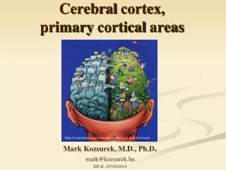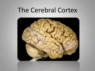
THE CEREBRAL CORTEX
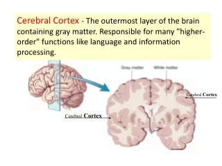
THE CEREBRAL CORTEX. Institute of Anatomy, 2nd Medical Faculty R. Druga. NEOCORTEX. Laminar pattern – 6 layers 10 – 20 billion neurons 95 % surface of the hemisphere. Pyramidal neurons Apical and basal dendrites Dendritic spines Excitatory (glutamate) Homogenous group 60 – 70 %.

THE CEREBRAL CORTEX
E N D
Presentation Transcript
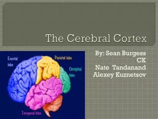
THE CEREBRAL CORTEX Institute of Anatomy, 2nd Medical Faculty R. Druga
NEOCORTEX • Laminar pattern – 6 layers • 10 – 20 billion neurons • 95 % surface of the hemisphere
Pyramidal neurons Apical and basal dendrites Dendritic spines Excitatory (glutamate) Homogenous group 60 – 70 % Non-pyramidal neurons Aspiny Heterogenous group Inhibitory (GABA) 30 – 40 % NEOCORTEX,types of neurons
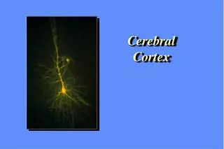
Pyramidal neurons Layer V. M I Golgi impregnation
Projection neurons, excitatory, glutamate Long axons Local circuit neurons, inhibitory, GABA Interneurons, short axons
My investigations showed that the functional superiority of the human brain is intimately bound up with the prodigious abundance and unusual wealth of forms of the so-called neurons with the short axons. S. R. y Cajal: Recuerdos de mi vida. 1917. Interneurons are butterflies of the soul. S.R. y Cajal 1923
Characteristics of layers • I.. Molecular layer – local inhibitory interneurons • II. External granular – association neurons • III. External pyramidal – commissural neurons • IV. Internal granular – receives thalamocortical projections • V. Internal pyramidal – projecting neurons (basal ganglia, brain stem, spinal cord • VI. Multiform layer – corticothalamic neurons
K.Brodmann, 1907, 1911 11 regions 52 areas
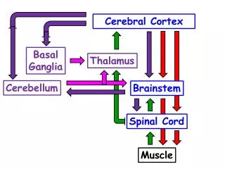
Afferent neocortical connections • Thalamic nuclei (thalamocortical fibers) • Amygdala • Claustrum • Nc. basalis (Meynert)-cholinergic system • Hypothalamus • Raphe nuclei (serotonin) • Locus coeruleus (noradrenalin) • Subst. Nigra (VTA) - dopamin
Excitatory connections in the neocortex • Layer 4 – termination of thalamocortical projections • Layer 4 – projects to layer 3 • Layer 3 – projects to layer 5
Efferent neocortical connections • Thalamic nuclei • Basal ganglia (striatum, amygdala, claustrum) • Brain stem (pretectal area, tectum, nc. ruber, RF, nuclei of cranial nerves, pontine ncc., nc. gracilis, nc. cuneatus) • Spinal cord ( corticospinal pathway, interneurons, motoneurons)
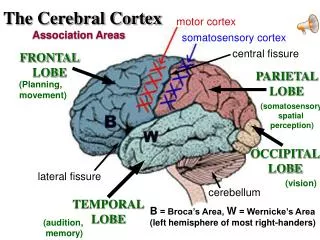
Motor cortical area • G. Fritsch and E. Hitzig (1870) demonstrated that electrical stimulation of the dog´s frontal lobe results in contralateral muscular contractions (movements)
Primary motor area M I • Precentral gyrus, area 4 • Part of the cortex from which movements are easily produced by electrical stimulation • Motor homunculus (overrepresentation muscles of the thumb, hand, face, tongue, somatotopic representation) • Afferents : S I, thalamic VL • Efferents : basal ganglia, thalamus, (VL) RF, superior colliculus, nc. ruber, RF, pontine ncc., spinal cort • Control of distal muscles • Damage produces paralysis of contralateral muscles (namely upper limb, tongue, facial muscles)
Premotor area, PM • Area 6 • Somatotopic representation of the body musculature, less precisely organized • Efferents – M I, basal ganglia, RF, Spinal cord (influences paravertebral and proximal limb musculature) • Afferents – thalamic VA (basal ganglia), S I, • Preparation to move
Supplementary motor area • Area 6, medial surface of the hemisphere • Somatotopic organization,less precisely organized • Afferents – thalamic VA (basal ganglia), parietal cortex • Efferents – MI, Basal ganglia, RF, Spinal cord • Area is involved in organizing and planning the sequence of muscle activation
Somatosensory area S I • Postcentral gyrus • Areas 3a, 3b, 1, 2 • Afferents : VPL, VPM • Efferents : M I, thalamus (VPL, VPM), pontine ncc., nuclei of cranial nerves (V.), spinal cord • 3a – signals from muscle spindles • 3b – cutaneous receptors • 2 – joint receptors • 1 – all modalities
LANGUAGE AREAS Broca: patient losses the ability to speak, produces single words, or syllables. Understanding of language is preserved. Often combined with agraphia. Wernicke : sensory or receptive aphasia, spontaneous speech is fluent, but sounds are often put together into meaningless words – „ word salad „. Often combined with alexia – the inability to read.
Auditory cortex • Area 41 • Afferents – auditory pathway (thalamic medial geniculate body) • Efferents – thalamus (medial geniculate body), inferior colliculus, associative cortical areas (what and where paths)
Visual cortex • Area 17, granular cortex • Afferents – visual pathway, thalamic lateral geniculate body • Efferents – thalamus (lateral geniculate body), area 18, 19, parietal cortex, temporal cortex. • Dorsal stream – parietal cortex (where : rods, periphery of retina, area 7) • Ventral stream – temporal cortex (what- colors, form : cones, central area of retina, area 37, inferior. temporal cortex
Corpus callosum – transekce
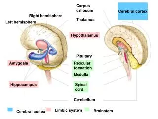
Cerebral cortex • All mammals depend on it • A man without a cortex is almost vegetable, speechless, sightless, senseless (D. Hubel and T. Wiesel 1979). • The cortex supports sensory perception, reasoning, planning and execution of behaviors