Functional localization of the cerebral cortex
420 likes | 786 Vues
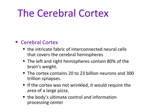
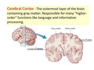
Functional localization of the cerebral cortex. Development. Three general areas have been proposed to define the different areas in the brain. General sensation: stimulation could elicit sensations of different kinds General motor: stimulation could cause muscular contractions.

Functional localization of the cerebral cortex
E N D
Presentation Transcript
Development • Three general areas have been proposed to define the different areas in the brain. • General sensation: stimulation could elicit sensations of different kinds • General motor: stimulation could cause muscular contractions. • Association area: increasing as evolution proceeds
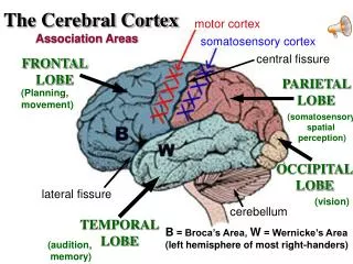
Parietal, Occipital, and temporal cortex • The first somesthetic area (general sensory area) • located at postcentral gyrus and posterior part of the paracentral lobule on the medial surface, in area 3, 1, 2 (It has some overlapping area with the precentral motor area)
First somesthetic area • Afferent fibers to the first somesthetic area are from ventral posterior nucleus of the thalamus (medial lemniscus, spinothalamic and trigeminothalamic tracts). The thalamocortical fibers traverse the internal capsule and medullary center (more later). The contralateral half of the body is represented as inverted. The pharyngeal region, tongue, and jaws are represented most ventral, followed by face, hand, arm, trunk, and the thigh. The area in cortical area is determined by the functional importance of the body part. Homunculus represents the body in proportion to its cortical map.
The second somesthetic area • located in the dorsal wall of the lateral sulcus, may extend into insula • receive fibers from intralaminar nuclei and posterior group of nuclei of thalamus. Afferent fibers to these nuclei come from reticular formation, spinothalamic and trigeminothalamic tracts. Provide less discriminative sensation, could be responsible for residual sensation after the first somesthetic sensory area is destroyed.
The somesthetic association area • located in the superior parietal lobule on the lateral surface and in the precuneus on the medial surface. In Brodmann’s area 5 and 7. Afferent fibers come from the first somesthetic area, providing comprehensive assessment of an object without visual aid.
Association area • Lesion of association area could lead to agnosia (inability to know familiar objects or persons). • Color Agnosia • Topographical Agnosia • Prosopagnosia
Color agnosia • a severely impaired ability to name and distinguish colors, even though basic color vision and brightness discrimination mechanisms are intact
Agnosia • Topographical agnosia patients are unable to use visual cues to guide them in a particular direction
Agnosia • Prosopagnosia refers to the inability to recognize faces despite good basic vision and intelligence, A person with a lesion causing prosopagnosia recognizes that a face is a face. However, a prosopagnosiac cannot distinguish one face from another
Agnosia • Lesion that destroys large portion of association cortex could lead to tactile agnosia and astereognosis. Patients are unable to tell different object by touch with eyes closed. Patients loss the ability to correlate texture, shape, size, and weight of the object with previous experience.
Astereognosis • One kind of loss of astereognosis is unawareness of spatial relations of parts of the body. Serve form (cortical neglect): which patient ignores or denies the existence of contralateral body.
Autotopagnosia • The word "auto" means self, and top is from topography, referring to the shape of the one's own body. An example will illustrate: A doctor asks a patient suffering from autotopagnosia, "show me your right hand." The patient responds, "Well, I did see it, it was around here somewhere. I guess I left it down in the cafeteria when I had lunch." The doctor replies, "all right, could you please point to your eyes?" The patient says, "oh, um, geez, oh yeea, they're over there," and points to a wall of the room. The doctor continues, "now, would you point to your nose?" The response is, "Oh, yeah, it's over there too."
Finger agnosia • There is one form of agnosia which is associated with the left parietal, temporal lesion, called finger agnosia. Finger agnosia is characterized by the lack of knowledge about one's hand. If a person suffering from finger agnosia is asked to draw a hand, the patient will draw a picture somewhat like this:
Vision • Visual area surrounds the calcarine sulcus on the medial surface of the occipital lobe (area 17, also called striate area). Fibers are from lateral geniculate body same side • Because of the partial crossing, left visual field is projected to right hemisphere and upper visual field is projected to lower wall of cortex (below calcarine line). Macula lutea (the yellow spot) project to posterior one third of the visual cortex. • Visual association cortex corresponds to area 18, 19. It receives info from area 17, function to relate the image to past visual experience. Lesion of area 18, 19 causes visual agnosia.
Visual association • Inferior temporal and lateral occipitotemporal gyri are another visual association area, where visual memories are stored, because stimulation of this area could cause visual hallucination. Bilateral destruction of this area causes apperceptive visual agnosia, one of which, prosopagnosia (unable to recognize other humans by their faces in spite of having good eye sight )
Hearing • Ventral wall of the lateral sulcus and the superior temporal gyrus (area 41, 42). • Medial geniculate body is the principal source of fibers that end in auditory cortex. Contralateral ear predominate, same side is also substantial • Auditory association cortex is at the floor of the lateral sulcus and posterior part of area 22 on the lateral surface of the superior temporal gyrus (also called Werbick’s area). Bilateral destruction of auditory association area causes auditory agnosia, fail to identify previous familiar sounds.
Taste • taste center is at the inferior end of the postcentral gyrus and extends into insula • Nerve impulses from taste buds reach gustatory nuclei, then ipsilaterally to the medial division of ventral posterior nucleus of the thalamus, then project to cortex by thalamocortical fibers
Olfaction • Most fbers end in limen insulae and uncus (area 34), some end in entorhinal cortex. Close to the taste center in brain
Vestibular representation • area unclear
Association cortex • close related to the primary sensory area and may be responsible for memory
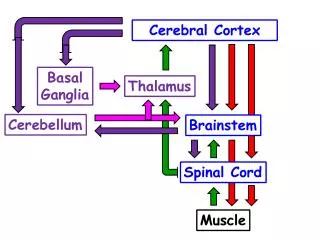
Frontal cortex • Motor area: primary motor area is located in the precentral gyrus (area 4) • input source are other motor areas in brain, like premotor area (area 6), somesthetic cortex, and posterior division of ventral lateral thalamic nucleus, which receives info from cerebellum. • Efferent fibers • originate from area 4 and area 6 and parietal lobe forming the corticospinal and corticobulbar tracts. The representation of body in primary motor area is inverted, similar to the sensory inputs in primary sensory cortex. • lesion of area 4 causes voluntray parelysis of the contrallateral side
Second and supplemental motor area • located at the part of area 6 on medial surface of the hemisphere and the cortex adjoining the anterior half of the cingulate sulcus. • Bilateral lesion could cause paralysis and akinetic mutism.
Premotor area • Coincides with area 6 and anterior to primary motor area • direct contribution to the pyramidal and other descending motor pathways • lesion in premotor area could lead to apraxia, loss of the ability to do simple or routine acts in the absence of paralysis. When writing is also involved, agraphia.
Prefrontal cortex • Envelop the frontal pole and corresponds to area 9, 10, 11, and 12. Only well developed in primates especially in humans. It has extensive connection through the medullary center to parietal, occipital and temporal lobes. Gaining access to current sensory information and the repository of data from past experience. • Bilateral prefrontal leukotomy (lobotomy) causes personality changes (rude, inconsiderate, rash, reckless, etc) but not suffering any severe pain, depression, or anxiety. • Disorders follow bilateral lesion to the prefrontal area, acquired sociopathy.
Language center • Two critical areas • 1. Receptive language area (sensory language area) consists auditory association area (Wernicke’s area) and the adjacent parietal lobe. • 2. Expressive speech center (Broca’s area, motor speech center) opercular and triangular portion of inferior frontal gyrus (area 44, 45)
Language • The two language centers communicate with each other by superior longitudinal fasciculus • These two language centers are located on the left side of the hemisphere which is the dominant hemisphere for most people in term of language.
Aphasia • Receptive aphasia: • Fluent but meaningless speech and severe impairment of the ability understand spoken or written words. Auditory and visual comprehension, naming objects and repetition of sentence are all affected. • Expressive aphasia: • Damage to Broca's area. OK comprehension but distorted speech (talking nonsense, patients are aware of this) whereas receptive aphasia patients are not aware of he/she is speaking nonsense.
A video clip about language • http://www.learner.org/resources/series142.html?pop=yes&vodid=185466&pid=1580#
Hemisphere Dominance • Because two hemispheres are connected by corpus callosum. Therefore, there are bilateral cortical memories for previous experiences.
Left hemisphere function • In right handed and most of the left handed people, language is the function of left brain. 75% of the population is right handed with language center on the left side of the brain and about 70% of the left handed people also with their language center on the left side.
Right hemisphere function • right hemisphere is responsible for three dimensional image, spatial perception (supported by evidence that after commissurotomy, removal of corpus collasum, left hand is more efficient handling tasks like arrange blocks, copying drawings). Right hemisphere may also be responsible for singing, and playing musical instruments. Lesion to the posterior part of the superior temporal gyrus causes amusia. • The right cerebral cortex on both side of lateral sulcus is necessary for prosody (verbal communication is intact, though), combination of tones, cadences, emphasis. Lesion to this area causes aprosodia (monotonous speech)