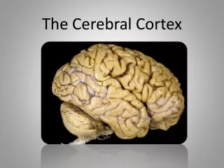
Cerebral cortex , primary cortical areas
281 likes | 597 Vues
Explore the structure and functions of key areas in the brain mantle, including Paleocortex, Archicortex, and Neocortex. Discover the evolution and organization of the cerebrum while diving into the classification and roles of neuronal cells.

Cerebral cortex , primary cortical areas
E N D
Presentation Transcript
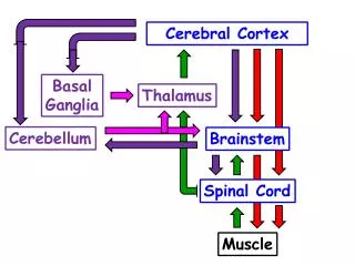
Cerebralcortex, primarycorticalareas Mark Kozsurek, M.D., Ph.D. mark@kozsurek.hu ED II., 07/10/2014 http://controlmind.info/human-brain/right-and-left-brain
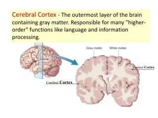
Ontogenesis of thecerebralcortex In a broader sense the pallium (or brain mantle) is the wall of the telencephalic vesicles and consists of both gray and white matters. As the telencephalon grows, it divides into paleopallium, archipallium and neopallium. The gray matter of these parts contributing to the hemispheric surfaces are summarized as paleocortex, archicortex and neocortex, respectively. (Note, that this classification is a bit obsolete, as new concepts appear year-by-year, but many of these also disappear without being confirmed.)
Phylogenesis of thecerebralcortex During thephylogenesis of Mammalstheproportion of thepaleocortex and thearchicortexdecreased, whiletheneocortexbecamedominantoccupying almost thewholesurface of thebrain. CORTEX Allocortex Isocortex (less than 6 layers) 6 layers PaleocortexArchicortex Not the total number of the layers, but the number of neuronal layers matters!
Paleocortex:theoldestcorticalarea of thetelencephalonwhichcontains 3 to 5 layers of neuronalcellbodies. Paleocortexincludestheolfactorybulb, olfactorytubercle (approx. attheanteriorperforatedarea) and thepiriformcortex (approx. theuncus and theanterior part of theparahippocampalgyrus). Allthosecortical and non-corticalareaswhicharerelatedtothesense of smellaresummarizedastherhinencephalonorolfactorybrain. Archicortex:constitutedby 3 to 4 layers of neurons and includesthehippocampusandrelatedstructures (dentateandfasciolargyri, indusiumgriseum). Neocortex:occupiesapprox. 90% of thetotalcerebralhemispherialsurface. 6 layers of neuronalcellbodiesarepresent.
1. Paleocortex http://www.nature.com/nrneurol/journal/v8/n6/full/nrneurol.2012.80.html http://www.the-scientist.com/?articles.view/articleNo/37607/title/Scent-Sorting/
2. Archicortex What you see in the microscope and the neuronal connections in the background...
GABAergic inhibitory neurons: 1. stellate cells (axo-dendritic) 2. basket cells (axo-somatic) 3. chandelier cells (axo-axonic) fimbria CA3 2. 3. mossy fibre granule cells in the dentate gyrus CA2 Schaffer’s collateral perforant path from entorhinal cortex pyramidal cells 1. CA1
CA3 fimbria mossy fibre CA2 str. moleculare Schaffer’s collaterals str. lacunosum str. radiatum perforant path In CA3 a str. lucidum containing mossy fibres is also present between str. pyramidale and radiatum. str. pyramidale str. oriens CA1 alveus
Molecular layer • Outer granule cell layer • Outer pyramidal layer • Inner granule cell layer • Inner pyramidal layer • Plexiform layer 3. Neocortex
PC: pyramidal cell, SSC: spiny stellate cell, BPC: bipolar cell, DBC: double bouquet cell, ChC: chandelier cell, MC: Martinotti cell, LBC: large basket cell, SBC: small basket cell, NBC: nested basket cell, NGC: neurogliaform cell, BTC: bitufted cell (dendrites in red and axons in blue) There are much more types of neurons in the neocortex than it is suggested by silver-impregnated specimens. And which neuron should be considered as a separate group?
The most characteristic cell type of the cerebral cortex: the pyramidal cell. Both its apical and basal dendrites posses spines and the axon arrises from the base of the conical cell body. http://cercor.oxfordjournals.org/content/15/6/802/F1.expansion
Functionalunits of thecortex:corticalcolumns • each column is approx. 200-300 µm wide and 2.5-3.0 mm tall (as thick as the cortex) • each cortical column contains approx. 5000 neurons and altogether 2 million such moduls constitute the human cortex (1000 columns in rats, 1 million in monkeys) • neurons predominantly contact other neurons within the same column (vertical communication) • each modul communicates with 100-200 other columns
Specificafferentsfromsensoryorgansthroughthethalamusmainlyterminateonspiny stellateneuronsinlaminaIV. Theiraxonsascendintolamina I and synapsewiththeapical dendrites of pyramidalcellsrepresentingthe output of thecorticalmodule. Corticocorticalafferentsreach almost allthelayers of thecorticalcolumns and synapsedirectlywithpyramidalcells. Importance of inhibitoryinter-neuronsincreasesclosetothemarginsofcorticalcolumnsastheyinhibitlateralspreadingofneuronalactivity, thus, isolateadjacentcolumnsfromoneother.
Different inhibitory interneurons terminate on distinct parts of pyramidal cells.
Connectionsamongcorticalmodules • Thalamocortical (specific) inputs terminate in lamina IV and make this layer thick in sensory cortical areas. • Corticocortical connections arise from lamina III pyramidal cells and terminate with extensively branching endings along the whole thickness of the target column. • Long descending motor pathways are mainly constituted by the axons of lamina V pyramidal cells. This is why this layer dominates in motor cortical areas. • Lamina VI sends axons back to the thalamus (corticothalamic projection). • Inhibitory neurons block lateral spreading of electric neuronal activity.
granular cortex / koniocortex agranular cortex
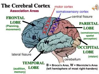
Corticalfields of Brodman • Brodmann, 1909 • 52 areas of the neocortex have been distinguished according to morphological parameters such as the thickness of layers, types and densities of different cells, etc. • Later became obvious that cortical fields described by Brodman are not only morphologicaly but also functionally distinct and have their own roles in different processes.
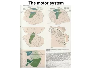
Motor cortical fields primary motor area (Br4): Betz cells, pyramidal tract premotor area (dorsolateral Br6) supplementary motor area (medial Br6) frontal eye field (Br8): voluntary eye movements Broca’s motor speach center (Br 44, 45) Sensory cortical fields primary somatosensory area (Br 3, 1, 2)
Visual cortical fields primary visual cortex (Br 17, striate area) termination of optic radiation: Gennari’s line secondary visual cortex (Br 18) tertiary visual cortex (Br 19) Auditory cortical fields primary auditory cortex (Br 41) secondary auditory cortex (Br 42) Wernicke’s speach area (Br 22, 39?, 40?)
As a rule of thumb: Primary cortical areas: site of origin of descending motor pathways and sites of termination of ascending sensory pathways. Secondary, tertiary cortical areas: association areas, information processing, memory (visual, auditory, etc.) Note the large extension of cognition- and vision-related areas!
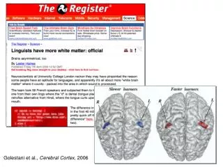
Hemisphericasymmetry – languageareas • Duetothedecussation of largeascendingsensory and denscending motor pathwayshemispheresfeel and movetheotherside of the body, butinspite of this, thetwohemispheresarenotcompletlysymmetrical. • Dominant:verbal, logical, abstract, analyzing, planning, expressing • Subdominant:non-verbal, workingwithimages, synthesizing, sensing • Right-handedpeople: • 96%: languageareasinthelefthemisphere. • Left-handedpeople: • 70%: ontheleft, • 15%: onthe right, • 15%: onbothsides.
Where are Wernicke's and Broca's Language Areas? Superimposition of various definitions of Wernicke's and Broca's areas on a standard anatomical drawing of the human left hemisphere.