
SlideServe
Menu
Préférences
© SlideServe · v2026

Somatic Sensory Pathways

isha avatar
14 diapositives · 1.1K Views · Aug 30, 2014

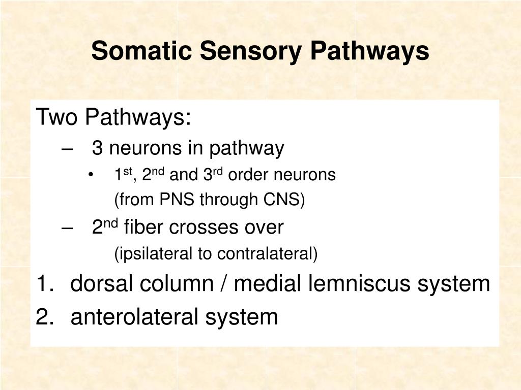
Somatic Sensory Pathways. Two Pathways: 3 neurons in pathway 1 st , 2 nd and 3 rd order neurons (from PNS through CNS) 2 nd fiber crosses over (ipsilateral to contralateral) dorsal column / medial lemniscus system anterolateral system. Fig. 7- 2 Ganong. Somatic Sensory Pathways.
Somatic Sensory Pathways - slide 1
01 / 14Somatic Sensory Pathways
1 / 17

Télécharger la présentation

Somatic Sensory Pathways
Une image / un lien ci-dessous est fourni (tel quel) pour télécharger la présentation

Politique de téléchargement : le contenu du site vous est fourni TEL QUEL pour votre information et votre usage personnel ; il ne peut être vendu, concédé sous licence ni partagé sur d’autres sites sans l’accord de son auteur.Le contenu vous est fourni TEL QUEL, pour votre information et votre usage personnel uniquement. Téléchargez la présentation en cliquant sur ce lien. Lors du téléchargement, si vous ne parvenez pas à télécharger une présentation, l’éditeur a peut-être supprimé le fichier de son serveur.Pendant le téléchargement, si vous ne parvenez pas à obtenir une présentation, le fichier a peut-être été supprimé par l’éditeur.

F I N

Plus de contenu associé

Transcription de la présentation
  1. Somatic Sensory Pathways Two Pathways: • 3 neurons in pathway • 1st, 2nd and 3rd order neurons (from PNS through CNS) • 2nd fiber crosses over (ipsilateral to contralateral) • dorsal column / medial lemniscus system • anterolateral system

  2. Fig. 7- 2 Ganong Somatic Sensory Pathways Fig. 13.4

  3. Dorsal Column / Medial Lemniscus System • Signals ascend spinal cord via the dorsal (white) columns. • large myelinated fibers (types I and II) • fine touch and proprioception • rapid transmission • high degree of spatial orientation Fig. 13.5

  4. Posterior Column / Medial Lemniscus System • 1st order sensory fibers ascend ipsilaterally in dorsal (posterior) columns to the medulla, and synapse in the medulla. • 2nd order fibers cross over in the medulla and travel through the brain stem via the medial lemniscus to the thalamus. • 3rd order fibers project to the somatosensory areas of the cerebral cortex. • post central gyrus of the parietal lobe

  5. Anterolateral System • Signals ascend the spinal cord via the anterior and lateral spinothalamic tracts. • small myelinated and unmyelinated fibers (types III and IV) • crude touch, pain, temperature • slow transmission • high volume of information • low spatial orientation Fig. 13.5

  6. Anterolateral System • 1st order sensory neurons enter spinal cord and synapse in the posterior horn. • 2nd order fibers cross over in the spinal cord, and ascend spinal cord to the thalamus via the anterior (ventral) and lateral spinothalamic tracts. • 3rd order fibers project to the somatosensory areas of the cerebral cortex. • post central gyrus of the parietal lobe

  7. Parietal LobePost Central Gyrus • homunculus • visual representation of the human body on the cerebral cortex • Because of crossover, the left side of the body projects to the right side of the cerebrum and vice versa. Fig. 14.21

  8. Somatic Sensation • Perception depends less on the source of the signal and more on what part of the brain is stimulated. • 2 examples from pain perception • phantom pain • referred pain

  9. Phantom Pain • Amputees feel pain in their (missing) limbs. • filling of cerebral cortical “vacuum” by other afferent signals? • activation of cut nerve endings?

  10. Referred Pain • Pain from viscera is perceived as coming from the skin. • e.g., heart attack: pain in left arm and shoulder • related to dermatomes Fig. 7-7 Ganong Fig. 16.4

  11. Regulation of Pain Sensation • Gate Theory • In the dorsal horn, the synapse between the first and second order neuron is a gateway for pain. • Pain may be blocked by “closing” this gate. • counter irritants • brain’s analgesia system: one of its sites of action gate Fig. 16.5 • collateral from touch receptor Fig. 7-2 Ganong

  12. Sensory Collaterals to the Brain Stem Stimulate the Reticular Activating System Fig. 16.3

  13. Subconscious Proprioception • via spinocerebellar pathways Fig. 13.4

  14. Video AssignmentThe Brain – Parts 1 and 2 • Part 1: The Enlightened Machine • Part 2: Vision and Movement • DVD and VHS versions are available. • Each part lasts about an hour. • See p.13 of Lecture Outlines for checkout information. • As you watch the videos, pay attention to who has what diseases.