Download
1 / 67
730 likes | 1.39k Vues
Neural Integration The sensory pathways Chapter 15. Afferent Division of the Nervous System. Receptors Sensory neurons Sensory pathways. Afferent Division – location in CNS. Somatic Sensory info Sensory cortex of cerebrum Cerebellum Visceral Sensory info Reflex centers in brainstem

E N D
Afferent Division of the Nervous System • Receptors • Sensory neurons • Sensory pathways
Afferent Division – location in CNS • Somatic Sensory info • Sensory cortex of cerebrum • Cerebellum • Visceral Sensory info • Reflex centers in brainstem • Reflex centers in diencephalon
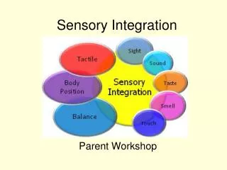
The somatic sensory system • Sensory stimuli that reach the conscious level of perception • Specialized cells that monitor specific conditions in the body or external environment • General Senses: • Temp, pain, touch, pressure, vibration, proprioception • Simple receptors located anywhere on body • Special Senses: • Are located in sense organs such as the eye or ear • Olfaction, vision, gustation, hearing, equilibrium • Complex receptors located in specialized sense organs
General Properties: Sensory Division Table 10-1 (1 of 2)
Sensory Pathways – from sensation to perception • Stimulus as physical energy sensory receptor • Receptor acts as a transducer • Intracellular signal usually change in membrane potential • Stimulus threshold action potential to CNS • Integration in CNS cerebral cortex or acted on subconsciously
Sensory Receptors • Transduction – conversion of environmental stimulus into action potential by sensory receptor • Receptors specific for particular type of stimulus • Specificity is due to structure of receptor
From Sensation to Perception • A stimulus is a change in the environment that is detected by a receptor • Sensation: the awareness of changes in the internal and external environment • Perception: the conscious interpretation of those stimuli
Classification by Location • Exteroceptors • Respond to stimuli arising outside the body • Receptors in the skin for touch, pressure, pain, and temperature • Most special sense organs • Interoceptors (visceroceptors) • Respond to stimuli arising in internal viscera and blood vessels • Sensitive to chemical changes, tissue stretch, and temperature changes
Classification by Location • Proprioceptors • Respond to stretch in skeletal muscles, tendons, joints, ligaments, and connective tissue coverings of bones and muscles • Inform the brain of one’s movements
Four types of General Sensory Receptors • Pain: nociceptor • Temperature: thermoreceptor • Physical: mechanoreceptor • Chemicals: chemoreceptors • All can be found in both somatic (exteroceptors) and visceral (interoceptors) locations except: • Proprioceptors (a mechanoreceptor) are somatic only • report the positions of skeletal muscles and joints
Pain Receptors: Nociceptors • (noci = harm) sensitive to pain-causing stimuli (e.g. extreme heat or cold, excessive pressure, inflammatory chemicals) • Free nerve ending • Mode of Action: • Injured cells release arachidonic acid • Arachidonic acid is converted into prostaglandins by the interstitial enzyme cyclo-oxygenase • Prostaglandins activate nociceptors • Many pain medications like aspirin function to inhibit cyclo-oxygenase • Pain levels are modulated by endorphins which inhibit CNS function
Thermoreceptors • Detect temperature • Found in skin, skeletal muscle, liver, and hypothalamus • Consist of free nerve endings • Phasic receptors that adapt easily • Cold response are more superficial and receptors that respond to heat – deeper • Temperature out of the range of the thermoreceptors will activate nociceptors
Mechanoreceptors • Detect membrane distortion • Three receptor types: • Tactile Receptors • Proprioceptors • Baroreceptors
Mechanoreceptors - Tactile Receptors • Detect touch, pressure and vibration on skin • Detect hair movement • Detect fine touch • Detect deep pressure • respond to itch (respond among other to histamine) and light touch (detect changes in shape like bending)
Mechanoreceptors - Proprioceptors • Detect positions of joints and muscles • Muscle spindles • Modified skeletal muscle cell • Monitor skeletal muscle length • Golgi tendon organs • Dendrites around collagen fibers at the muscle-tendon junction • Monitor skeletal muscle tension • Joint capsule receptors • - Monitor pressure, tension and movement in the joint
Mechanoreceptors - Baroreceptors • Detect pressure changes • Found in elastic tissue of blood vessels and organs of digestive, reproductive and urinary tracts
Chemoreceptors • Detect change in concentration of specific chemicals or compounds • pH, CO2, sodium etc. • Found in respiratory centers of the brain and in large arteries
Sensory Receptors Table 10-2
Processing of the sensory information • Levels of neural integration in sensory systems: • Receptor level — the sensor receptors • Circuit level — ascending pathways in the CNS • Perceptual level — neuronal circuits in the cerebral cortex
Processing at the Receptor Level Perceptual level(processing in cortical sensory centers) 3 Motor cortex Somatosensory cortex Thalamus Reticular formation Cerebellum Pons Medulla Circuit level (processing in ascending pathways) 2 Spinal cord Free nerve endings (pain, cold, warmth) Muscle spindle Receptor level (sensory reception and transmission to CNS) 1 Joint kinesthetic receptor Figure 13.2
Processing at the Receptor Level • The receptor must have specificity for the stimulus energy (as previously discussed) • The receptor’s receptive field must be stimulated • The stimulus need to be converted to a nerve impulse • Receptors have different levels of adaptation • Information is encoded in the frequency of the stimuli – the greater the frequency, the stronger is the stimulus.
The stimulation of the receptive field affects the discharge of the sensory neurons • The receptive field is the a specific physical area that, when stimulated, affect the discharge of the stimulus. • Most receptive fields activation will result in message sending – excitatory receptive field • Sensory receptors in the CNS can have inhibitory receptive field (example: vision fields to determine borders). • Sensory neurons of neighboring receptive field may exhibit • Convergence many sub-threshold stimuli to sum in the postsynaptic neuron • Overlapping with another receptor’s receptive field – sending 2 sensations from the same area (pressure and pain) • The smaller the receptive field the greater the ability of the brain to localize the site
Sensory Neurons: Two-Point Discrimination (a) Compass with pointsseparated by 20 mm • convergenceTwo-point discrimination Skin surface Primarysensoryneurons Secondarysensoryneurons One signal goes to the brain. Figure 10-3a
(b) Compass with pointsseparated by 20 mm Skin surface Primarysensoryneurons Secondarysensoryneurons Two signals go to the brain. Sensory Neurons: Two-Point Discrimination - overlapping Figure 10-3b
Receptive Fields of Sensory Neurons - overlapping Primary sensoryneurons The primary sensory neuronsconverge on one secondarysensory neuron. Information from thesecondary receptivefield goes to the brain. Secondarysensoryneuron The receptive fields of three primary sensory neuronsoverlap to form one large secondary receptive field. SECTION THROUGH SPINAL CORD Figure 10-2
Properties of Stimulus: Location • Lateral inhibition enhances contrast and makes a stimulus easier to perceive Stimulus Stimulus Pin Skin A B C Frequency of action potentials Tonic level Primary neuronresponse is proportionalto stimulus strength. Primarysensoryneurons Pathway closest tothe stimulus inhibitsneighbors. Secondaryneurons A B C Frequency of action potentials Inhibition of lateralneurons enhancesperception of stimulus. Tonic level Tertiaryneurons A B C Figure 10-6
Transduction allows sensory receptors to respond to stimuli – converting sensation into a nerve impulse • Sensory transduction – the process that enables a sensory receptor to respond to a stimulus. • The sensory transduction induces a receptor potential in the peripheral terminal of the sensory neuron • A receptor potential is a depolarization event that if brings the membrane to a threshold, will become a nerve impulse (AP) • The conversion from receptor potential to AP happens in the trigger zonethat can be in the first node of Ranvier. • In some cases, the peripheral terminal is a separate sensory cell (ex. Photo receptors). In this case there is an involvement of a synapse and NT
Receptors adaptation • The duration of a stimulus is coded by duration of action potentials. • A longer stimulus generates longer series of APs. • If a stimulus persists, some receptors adapt or stop responding • There are 2 classes of receptors according to how they adapt: • Tonic receptors – slowly adapting – they fire rapidly when first activated, than they slow and maintain firing as long as the stimulus is present (baroreceptors, proprioceptors) • Phasic receptors – rapidly adapting receptors – rapidly firing when first activated but stop firing if the strength of stimulus remains constant • This type of reaction allows the body to ignore information thatwas evaluated and found not to be a threat to homeostasis (smell)
Tonic Receptors • Always active • Signal at different rate when stimulated • Monitor background levels Figure 10-8a
Phasic Receptors • Activated by stimulus • Become active for a short time whenever a change occurs • Monitor intensity and rate of change of stimulus Figure 10-8b
Receptors adaptation • The mechanisms for receptors’ adaptation depends on the receptors: • Potassium channels in the receptor’s membrane open causing the membrane repolarization • Sodium channels inactivated stopping depolarization • Accessory structure may contribute to decrease sensitivity (muscle in the ear contract and limit the movement of the auditory oscicles)
Processing at the circuit Level Perceptual level(processing in cortical sensory centers) 3 Motor cortex Somatosensory cortex Thalamus Reticular formation Cerebellum Pons Medulla Circuit level (processing in ascending pathways) 2 Spinal cord Free nerve endings (pain, cold, warmth) Muscle spindle Receptor level (sensory reception and transmission to CNS) 1 Joint kinesthetic receptor Figure 13.2
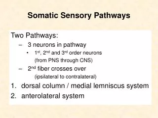
Processing at the circuit Level • A sensory pathway is a set of neurons arranged in series. • The circuit level role is to deliver the impulses to the appropriate region in the cerebral cortex. • The ascending tract typically consists of 3 neurons • First order neurons • cell bodies in a ganglion (dorsal or cranial) • Impulses from skin and proprioceptors to spinal cord or brain stem to a 2nd order neuron • Second order neuron • In the dorsal horn of the spinal cord or in the medulary nuclei • Transmit impulses to thalamus or cerebellum • Third order neurons • Cell bodies in the thalamus (no 3rd-order neurons in the cerebellum) • Transmit signals to the somatosensory cortex of the cerebrum
Pathways for somatic perception • Receptors for the somatic sensations are found both in the skin and viscera • Receptor activation triggers AP in the 1st order neuron • In the spinal cord, sensory neurons synapse with interneurons – 2nd order neurons • All 2nd order neurons cross overat some point (sensations are being integrated in the opposite side) • The synapse between the 2nd and the 3rd happens in the thalamus • The axons of the 3rd order neurons project to the appropriate somatosensory area in the cerebral cortex
Processing at the circuit Level • Impulses ascend in : • Non specific pathway that in general transmit pain, temperature and touch • Give branches to reticular formation and thalamus on the way up • Sends general information that is also involved in emotional aspects of perception • Specific ascending pathways involve in more precise aspect of sensation
Thalamic Function • The thalamus is the “gateway to the cerebral cortex” • Major relay station for mostsensory impulses that arrive to the primary sensory areas in the cerebral cortex: • taste, smell, hearing, equilibrium, vision, touch, pain, pressure, temperature • Contributes to motor functions by transmitting information from the cerebellum and basal ganglia to the cerebral primary motor area • Connects areas of the cerebrum • Impulses of similar function are sorted out, edited, and relayed as a group
3 major somatosensory pathways –1) spinothalamic pathway • Conscious sensation of poorly localized sensations • Anterior spinothalamic tracts – crude touch and pressure • Lateral spinothalamic tracts – pain and temperature • 1st order neurons synapse with the 2nd in the posterior gray horn at the level of entrance • The 2nd cross before ascending to the thalamus • 3rd order synapse at the level of the primary somatosensory cortex
3 major somatosensory pathways - 2) Posterior column pathway • Sensation of precise touch, vibration and proprioception • Includes • Left and right fasciculus gracilis (inferior part of the body) • Left and right fasciculus cuneatus (superior part of the body) • First order neurons enter the CNS at the dorsal roots and the sensory roots of cranial nerves. • Synapse with 2nd order in the medulla • 2nd order neurons cross over in the brain stem • 3rd order in the thalamus where the stimuli are sorted by the nature of stimulus and the region of body involved
3 major somatosensory pathways – 3) The spinocerebellar pathway • Information about muscle, tendon and joint position from the spine to the cerebellum • This information is subconscious • 1st order neurons synapse in the dorsal horn • 2nd order neurons ascend via anterior and posterior spinocerebellar tracts to the cerebellar cortex • Used to coordinate movements • In this pathway there is no 3rd order neuron
Somatic Senses Pathways Sensations are perceivedin the primary somaticsensory cortex. 4 4 Sensory pathwayssynapse in the thalamus. 3 3 THALAMUS MEDULLA 2 Fine touch, vibration,and proprioceptionpathways cross themidline in the medulla. 2 Fine touch,proprioception,vibration KEY Pain, temperature, andcoarse touch cross themidline in the spinal cord. 1 1 Nociception,temperature,coarse touch Primary sensory neuron Secondary sensory neuron Tertiary neuron Figure 10-9, steps 1–4 SPINAL CORD
Processing at the Perceptual Level Perceptual level(processing in cortical sensory centers) 3 Motor cortex Somatosensory cortex Thalamus Reticular formation Cerebellum Pons Medulla Circuit level (processing in ascending pathways) 2 Spinal cord Free nerve endings (pain, cold, warmth) Muscle spindle Receptor level (sensory reception and transmission to CNS) 1 Joint kinesthetic receptor Figure 13.2