Download
1 / 63
630 likes | 874 Vues
D-Dimers: Clinical Utility in the Diagnosis of PE. Resident Grand Rounds November 19, 2002 Heather M. Powers, MD Wake Forest University Medical Center Department of Internal Medicine. Overview. Case presentation Clinical questions Epidemiology VTE D-dimers – pathophysiology Assays

E N D
D-Dimers:Clinical Utility in the Diagnosis of PE Resident Grand Rounds November 19, 2002 Heather M. Powers, MD Wake Forest University Medical Center Department of Internal Medicine
Overview • Case presentation • Clinical questions • Epidemiology VTE • D-dimers – pathophysiology • Assays • Evidence • Conclusions
Case Presentation • 45 yo obese WF presenting to ED • PMHx significant for asthma • Complaints of SOB and right sided chest pain x 1 day • Reports recent travel • Medication OCP’s • Soc HX nonsmoker, works at desk job • Fam Hx • PE: VVS 92% pulse ox RA • Lungs: few scattered wheezes, LE: no asymmetry • CV: RRR w/o m/r/g
Case (cont’d) Would a D-dimer be useful in ruling out PE in this patient with a low pretest probability without doing diagnostic imaging?
Clinical Questions • How sensitive and specific are D-dimers at detecting VTE? • In what patient population is it appropriate to use D-dimers in helping rule out VTE? • Can you safely withhold anticoagulation in patients with a negative D-dimer result? • Can D-dimers be used as the sole test to rule out VTE?
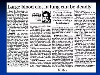
VTE • refers to spectrum of PE and DVT • Annual incidence of VTE • 125,000-400,000 cases/year (some as high as 2 million) • PE responsible for 200,00 deaths in US /year • preventable condition associated with substantial morbidity and mortality if untreated • Untreated 30% (23-87%) • Treated 2-8% • 65-90% of PE’s arise from LE
Age > 40 Immobilization Obesity h/o CVA Recent surgery (within the last 3 months) Estrogen use Lower extremity fractures Myocardial Infarction VTE - Risk Factors 40 years old CVA
CHF Varicose veins Pregnancy/postpartum Antiphospholipid Ab Prior history of VTE OCP’s Hypercoagulable state (i.e. Factor V Leiden) VTE - Risk Factors DVT PE FACTOR V CHF
Signs tachypnea (70%) rales (51%) tachycardia (30%) fourth heart sound (24%) accentuated P2 (23%) fever (14%) Symptoms dyspnea (73%) pleuritic pain (66%) cough (37%) hemoptysis (13%) Clinical Predictors of PE? • Caveat • frequency of these symptoms and signs similar to the 843 PIOPED cohort patients w/o PE
Diagnostic Dilemmas • History and physical exam • Objective tests • Gold standard tests • V/Q scanning • Empiric anticoagulation
What are D-Dimers? • degradation product from a specific region of cross-linked fibrin
What are D-Dimers? • Degradation product from a specific region of cross-linked fibrin • Sensitive markers for thrombosis but not specific • commonly elevated in patients with malignancy, infection, CHF, renal failure, SS crisis, and recent surgery • Levels elevated 8 fold after VTE compared with controls • Levels normalize within 15-20 days • Levels increase linearly with age
Effects of age on D-dimer performance % Age(yrs) Adapted from Righini et al, 2000
Relationship between D-dimer levels and Embolus Location • De Monye demonstrated that the accuracy of the D-dimer measurement to exclude PE depends strongly on embolus location • D-dimer may miss subsegmental PE’s Adapted from de Monye et al, Amer J of Resp Crit Care Med 2002
ELISA Microplate ELISA gold standard VIDAS ELISA with reported sensitivity 100% Typically more time consuming but newer tests more rapid Less specific Agglutination Second generation kits have higher sensitivity and are more clinically useful (i.e. SimpliRED) More rapid More specific Assays • Correlation between different assays is poor
Performance of various D-dimer assays in suspected PE Pts (n) PE (n) Sensitivity Specificity Classical ELISA Rapid ELISA Classical latex tests Microlatex WBL test Adapted from Task Force Report Eur Heart Journal, 2000
Evidence • Basic EBM concepts • Pretest probability • Likelihood ratios • Clinical Evidence
Formulating PTP • Pretest Probability • Probability that a patient has the disease before undergoing a test PTP + LR = posttest prob • LR > 10 or < 0.1 significantly changes post-test probability
Low <2 Moderate 2-6 High >6 Modified Wells CriteriaAdapted from Wells et al., 1998 Clinical FeaturesScore Clinical signs and symptoms of DVT 3.0 HR>100 beats/min 1.5 Immobilization (for >3 consecutive days) 1.5 Surgery in previous 4 weeks 1.5 Previous dx of DVT or PE 1.5 Hemoptysis 1.0 Cancer 1.0 PE as likely or more likely than any other diagnosis 3.0
ROC Curve for Plasma D-dimerELISA Adapted from Perrier in American Journal of Respiratory Critical Care Medicine, 1997
Bates et al, “A latex D-Dimer reliably excludes venous thromboembolism” 2001 • Question • Does the MDA- D-dimer assay have a high sensitivity and NPV for VTE and does it have the specificity to make the test clinically useful? • Design • Retrospective cohort study • Population • 595 (60% women) unselected outpatients with clinically suspected DVT (317) or PE (278) referred to 4 tertiary care centers in Canada
Bates et al • Methods • Pts assigned pretest probability of VTE based on previously validated model • Blood drawn for testing with MDA D-dimer assay (negative <0.5µg FEU/ml) • Evaluated with objective testing per consulting physician • Pts with low PTP and (–) D-dimer assay had no further testing • Anticoagulation held in all patients with (-) objective test results • Classified as VTE + or – according to the objective test results and followed for 3 mo.
Bates et al • Results • Prevalence of VTE was 19%(113/595) • 48 pts (17.3%) with suspected PE classified as PE+ • 34.5% (205) had a low PTP of VTE prevalence of VTE in low PTP group 5.9% and 16.9% in moderate PTP group • MDA D-dimer has 97% sensitivity and 99% NPV when low and moderate PTP groups combined • LR(-) was 0.07 in this subgroup of patients
Accuracy Indices of the D-dimer in patients with suspected VTE
Bates et al • Conclusions • First study to demonstrate that a D-Dimer assay has the potential to be used as the sole diagnostic test to exclude VTE • 96% sensitivity and 98% NPV for all comers with suspected VTE • NPV compares favorably with widely accepted diagnostic tests for VTE • D-dimer result of <0.5µg FEU/ml excludes VTE in patients with a low or moderate pretest probability (80% of population in this study) • Promising results but need further testing to determine safety of withholding anticoagulation in patients with low/mod pretest probability and (-) MDA D-dimer test
Strengths Specificity of this latex assay higher than ELISA Evaluated subgroup of pts with cancer with low/ mod PTP of VTE and found 97% sensitivity and 94% NPV Limitations Did not perform gold standard Results cannot be extrapolated to other assays Funded by Organon Teknika, manufacturer of MDA assay Bates et al
Ginsberg, et al “Sensitivity and specificity of a rapid whole blood assay for D-dimer in the diagnosis of pulmonary embolism” 1998 • Question • How accurate is a D-dimer assay in patients with suspected PE? Can a (-) D-dimer be of value in excluding PE in patients with low probability, non-diagnostic lung scans, or both? • Design • prospective cohort with blinded comparison of D-dimer assay results, V/Q scanning, and bilateral CUS with watchful waiting (3mo) • 4 tertiary care centers from Sept ‘93-May ’96
Ginsberg et al • Population • 1177 consecutive adults >18 yrs of age referred for suspected PE. • Exclusion criteria • Suspected UE DVT • No symptoms within previous 48hrs • Receipt of anticoagulation for 72hrs • Limited life expectancy • Contraindication for contrast media
Ginsberg et al • Methods • Classified as high, moderate, or low pretest probability. All patients underwent V/Q scanning and CUS within 24 hours of presentation • D-dimer tests using SimpliRED assay done concurrently with other diagnostic testing • All patients followed for 3 months
Ginsberg et al • Results • Overall prevalence of PE 17% (197) • No patient classified as PE (-) died of PE during follow up • For all patients, D-dimer assay had a sensitivity of 84.8% and a specificity of 68.4% • Subgroup analysis based on PTP • 703 classified as low PTP and 3.4% PE(+) • 521 had (-) D-dimer, 5 of which were PE(+) • LR (-) = 0.27
Ginsberg et al PE in low PTP Sens = 79% Spec = 76% NPV = 99.7% LR (-) = 0.27 D- Dimer
Ginsberg et al • Conclusions • D-dimer testing has a high sensitivity and moderate specificity for PE • D-dimers should not be used as a stand alone test • Not to be used to exclude PE in patients with mod/high pretest probability unless combined with other objective testing • Further testing and anticoagulant therapy may be with held in patients with a normal D-dimer if patients have a low pretest probability
Ginsberg et al • Limitations • Gold standard not performed • Results apply only to SimpliRED assay • Outpatients only • Did not give details of follow up data
Perrier A. et al “Non-invasive diagnosis of venous thromboembolism in outpatients”, 1999 • Design • Prospective cohort study with 3mo. follow up • Population • 918 consecutive patients presenting to ED or outpatient clinic with suspected PE or DVT • Methods • Clinical probability assigned based on RF, signs and sxs, likelihood of alternative diagnosis, bld gas and CXR results in case of PE
Perrier et al • Methods (cont’d) • Low, moderate, and high probability defined as 0-20%, 21-79%, and 80-100% respectively • Pts then entered algorithm • D-dimer testing (VIDAS ELISA) performed on all pts • D-dimer <500µg/L deemed to r/o VTE • Pts with (+) dimer results went further testing according to the algorithm • Pts followed at 3mo for determination of bleeding or thromboembolic events
Perrier et al • Primary outcome • Efficiency of their diagnostic strategy in terms of the number of patients in whom a definite diagnosis can be made without invasive workup
Perrier et al • Results • Prevalence of VTE was 23% and similar in both DVT and PE subgroups • D-dimer normal in 31% of cohort therefore excluding VTE • 2 FN tests resulting in NPV of 99.3% • D-dimer and ultrasound established diagnosis in 48% of cohort • Overall a noninvasive dx of VTE achieved in 99% of pts
Perrier et al • Prevalence of DVT and PE in low probability subgroup was 2% and 9% respectively • 10 of the 703 pts not on anticoagulation had confirmed events during 3 mo F/U • None of these 10 pts had a normal D-Dimer • Overall 3 month thromboembolic risk was 1.8%
Perrier et al PE / DVT Sens =99.1% Spec = 41.1% NPV = 99.3% LR (-) = .02 D-Dimer
Perrier et al • Conclusions • High sensitivity in all pts using VIDAS ELISA • Noninvasive workup feasible in a large proportion of patients • Low thromboembolic risk at 3 months • Large cohort and therefore easily applied to outpatient population presenting with suspected DVT or PE
Perrier et al • Limitations • Applies only to outpatient population • Gold standard not performed in all patients • Did not follow a clinical prediction rule
Wells et al, “ Excluding PE at the bedside without diagnostic imaging: Management of patients with suspected PE presenting to the ED by using a simple clinical model and D-dimer” 2001 • Question • How safe is using a clinical model combined with D-dimer assay to manage patients presenting to the ED with suspected PE? • Design • prospective cohort study • Population • 930 consecutive patients with suspected PE at 4 tertiary care ED in Canada from Sept 98-Sept 99
Wells et al • Methods: • Pretest probability of PE assigned using clinical model • SimpliRED D-dimer testing performed in all pts after determining PTP • Pts with low PTP and (-) D-dimer were considered PE (-) and had no further testing • All other pts had V/Q scans
Low <2 Moderate 2-6 High >6 Modified Wells CriteriaClinical Assessment for PE Clinical FeaturesScore Clinical signs and symptoms of DVT 3.0 HR>100 beats/min 1.5 Immobilization (for >3 consecutive days) 1.5 Surgery in previous 4 weeks 1.5 Previous dx of DVT or PE 1.5 Hemoptysis 1.0 Cancer 1.0 PE as likely or more likely than any other diagnosis 3.0