Download
1 / 32
320 likes | 711 Vues
Tissue of the teeth. Dr Jamal Naim PhD in Orthodontics. Periodontium (cont.). Sharpey's Fibers.

E N D
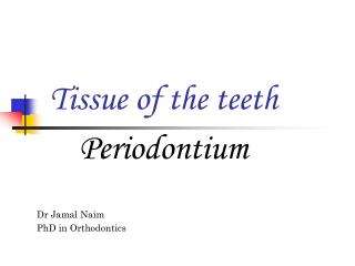
Tissue of the teeth Dr Jamal Naim PhD in Orthodontics Periodontium (cont.)
Sharpey's Fibers Sharpey's fibers always emerge from the cement in a straight line and continue across the periodontal interval and into the alveolar bone stresses are then always applied in the direction of their long axis.
Cementicles Cementicles are small-mineralized bodies, which may be found in the periodontal ligament. They may be attached to the cementum or the alveolar bone, or occur free in the periodontal ligament. When present, cementicles are generally found about all or most of the teeth. Cementicles may be formed by mineralization of degenerating epithelial rests or thrombosed vessels.
to bind the tooth to alveolar bone.
Age changes of cementum Hypercementosis: It is an abnormal thickening of cementum, may be diffuse or circumscribed. It may affect all teeth of the dentition, be confined to a single tooth, or even affect only Parts of one tooth. If the overgrowth improves the functional qualities of the cementum, it is termed a cementum hypertrophy. If the overgrowth occurs in nonfunctional teeth, it is termed hyperplasia (e.g. ostitis deformans paget).
Localized hypercementosis Generalized hypercementosis
Age changes of cementum In Localized hypertrophy prong like extension of cementum may be formed. This condition frequently is found in teeth that are exposed to great stress (ortho) (compensatory cementum). This extension of cementum provide a larger surface area for the attaching fibers; thus a firmer anchorage of the tooth to the surrounding alveolar bone is assured.
Age changes of cementum Localized hypercementosis may some times due periapical inflammation. Here the hyperplasia is circumscribed and surrounds the root like a cuff.
Age changes of cementum Although root resorption is not a part of the normal functional activity in the permanent dentition, most teeth show minute areas of resorption which may extend through the cementum and into the root dentin. The resorptive activity may have been initiated by trauma from occlusion, orthodontic forces or for unknown reasons. Generally, the defect is repaired by rapid deposition of cellular cementum once the initiating factor is removed.
Types of bone • Lamellar bone • compact bone • spongy bone • Woven bone • Bundle bone
Bone Trabeculae Bone Marrow Spaces
3. Bundle Bone It is referred to the bundles of principal fibers of either the periosteum or PDL continue into the bone as sharpeys fibers.
Alveolar bone The alveolar process is that bone containing the alveoli. It consists of: • an outer (lingual and buccal) cortical plate (compact bone) • A central spongiosa (spongous bone) • and • Alveolar bone (bone lining the alveolus), (bundle bone) The alveolar bone and the cortical plate meet at the alveolar crest (1.5 to 2 mm below the level of CEJ).
alveolar crest Cortical plate alveolar bone
Alveolar bone The alveolar bone is perforated by many foramina to allow blood and nerve supply to the teeth, so it is referred to cribriform plate Hirschfeld canal
Alveolar bone In X-ray the cribriform plate is referred to lamina dura.
Alveolar bone The bone directly lining the socket (inner aspect of the alveolar bone) is referred to bundle bone. In the alveolar bone we can found all histological forms of bone, but almost bundle bone. Embedded in the bundle bone are the collagen fiber bundles of the PDL, that provides attachment of the tooth to the alveolar bone.
Alveolar bone Collagen fibers
Alveolar bone Outer compact bone Spongiosa Collagen fibers
Alveolar bone Cementum Spongiosa Dentin PDL
Alveolar process The cortical plate of alveolar process consists of fine fibered lamellar bone It is generally thinner in the maxilla and thickest on the buccal aspect of the mandibular molars and premolars. Trabecular bone is absent in the region of frontal teeth, so that cortical plate and alveolar bone are fused together.