Download
1 / 83
870 likes | 1.17k Vues
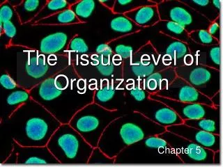
The Tissue Level of Organization. Chapter 5. What is a Tissue?. The human body is composed of trillions of cells There are approximately 200 cell types that make up the trillions of cells

E N D
The Tissue Level of Organization Chapter 5
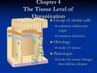
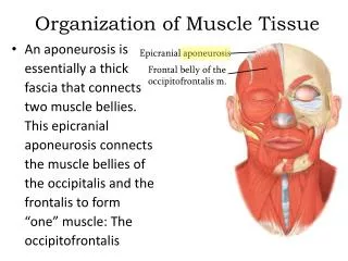
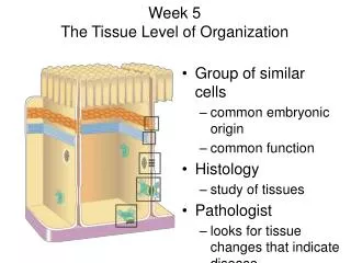
What is a Tissue? • The human body is composed of trillions of cells • There are approximately 200 cell types that make up the trillions of cells • These 200 cell types can be further grouped into 4 categories according to their roles – these 4 categories are called tissues • Tissues are groups of cells that function together to keep a body alive
Types of Tissues • Epithelial Tissue • Connective Tissue • Muscle Tissue • Neural Tissue
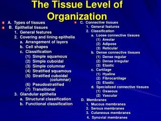
Epithelial Tissue Consist of 2 sub-categories: 1. Epithelia 2. Glands Epithelia are layers of epithelial cells that cover the internal or external surfaces of various organs, ducts, vessels, etc. Glands are structures whose cells produce fluid secretions. They are either attached to epithelia or made from epithelia
Characteristics of Epithelia • Cellularity – made up entirely of cells that are closely bound by tight junctions, gap junctions and desmosomes • Polarity – One end of the tissue usually faces an internal or external area of an organ (apical surface) and the other end is either attached to other tissues or a basement membrane called a basal lamina (basolateral surface) • Avascularity – Epithelia do not contain blood vessels – they get their oxygen and nutrients from adjacent cells. • Regeneration – Epithelial cells are constantly lost on the exposed surface – so they are constantly replaced by cell division of stem cells
Apical surfaces of internal passageways may have special extensions called microvilli POLARITY AND THE CONTROL OF PERMEABILITY
What are the Functions of Epithelial Tissue? • Provide Physical Protection – against abrasion, dehydration and destruction from chemical or biological agents • Control Permeability – some allow substances to enter / leave or pass through, other epithelial cells are quite impermeable • Provide Sensation – large sensory nerve supply • Produce Specialized Secretions – These epithelial cells are called gland cells (Cells of the thyroid, salivary glands, etc.) They use blood and interstitial fluid as carriers of the chemicals they release
Maintaining Epithelia Integrity I. Intercellular connections • CAMs (Cell Adhesion Molecules - proteins) • Intercellular cement (Proteoglycans, glycosaminoglycans such as Hyaluronan) • Cell Junctions • Tight Junctions – prevent passage of fluids and solutes (e.g. Stomach lumen) • Gap Junctions – proteins called connexons leave gaps for passage of various materials • Desmosomes – CAMs and proteoglycans link adjacent cell membranes with internal intermediate filaments– strong, resist stretching and pulling • Belt Desmosomes, Button Desmosomes and Hemidesmosomes (Between cell and basal lamina)
Acantholysis and Desmosomes Pemphigus • Pemphigus involves blistering of the outer (epidermal) layer of the skin and mucous membranes. It is an autoimmune disorder in which the immune system produces antibodies against specific proteins in the skin and mucous membrane. These antibodies produce a reaction that leads to a separation of epidermal cells (acantholysis). The antibodies usually develop against desmosome proteins. Loss of desmosomes causes epidermal cells to “Float away”
Maintaining Epithelia Integrity II. Attachment to the Basal Lamina Epidermal cells hold each other as well as the basal lamina which consists of 2 layers: Lamina lucida – consists of proteins secreted by adjacent cells – acts as a barrier that restricts movement of proteins Lamina Densa – contains bundles of proteins that gives the basal lamina strength Hemidesmosomes finally attach epithelial cells to the complete basal lamina
Maintaining Epithelia Integrity III. Epithelial Maintenance and Repair Stem cells or germinative cells within the epithelial tissue divide continually, to give rise to new epithelial cells to replace those that have died out due to exposure to toxic chemicals, pathogenic bacteria, abrasions, etc.
Simple Squamous • Thin, single layer of flat cells attached to basal membrane (lamina) • Substances pass through very easily • Forms walls of capillaries, lines other blood and lymph vessels, lines insides of certain organs • Lines alveoli of lungs • Easily damaged!
Simple Cuboidal • Single layer of cube-shaped cells attached to basal membrane • Covers ovaries, lines tubes and ducts of kidneys, certain glands such as pancreas, salivary glands and liver • Can be used to secrete glandular products or reabsorb materials such as water (in kidneys)
Simple Columnar • Single layer of elongated cells, attached to basal membrane • Cells may or may not be ciliated on their apical surface • Nonciliated simple columnar epithelia lines the uterus, parts of the GI tract and • Can secrete digestive juices • Because of thickness, they protect underlying tissues • Ciliated simple columnar epithelia lines the fallopian tubes, to move eggs • Some line intestines and have villi and microvilli to increase surface are for absorption
Pseudostratified Columnar • Appears stratified, but is not • Apical surface of cells have cilia • Line respiratory tract • Contain special cells called goblet cells that secrete mucous to trap dust and microorganisms
Stratified Squamous • Many layers of squamous cells, so relatively thick • Cells at apical surface are flattened and buildup a protein called keratin, which protects them from water damage, microorganisms, etc. Cell division occurs in deeper layers where cells are cuboidal or columnar • Upper layer of skin (keratinized)- tough, dead • Oral cavities, throat, esophagus, vagina, anal canal – all non-keratinized; upper layer cells are soft, alive.
Stratified Cuboidal • 2 or 3 layers of cuboidal epithelium • Line the ducts of salivary glands, mammary glands, sweat glands, etc. • Also lines immature or developing tubes in the male and female reproductive systems
Stratified Columnar • Cells at apical end tend to be columnar (elongated) whereas cells at the basal level are cuboidal in shape • Line the vas deferens, male urethra and areas of the pharynx
Transitional Epithelium • Changes in response to increased tension • Able to stretch; also prevents materials in the urethra from reentering the tissues • Lines inside of uterus, urinary bladder and urethra • When walls of these organs expand, the cells appear cuboidal • When walls stretch (distend), the cells appear flattened
Glandular Epithelium • Cells specialized to produce and secrete substances into ducts leading out of the gland • Glandular cells are actually embedded in and surrounded by columnar and cuboidal epithelial cells • Two types of glands: • Exocrine: secrete products into ducts that that open into and internal or external surface (goblet cells, salivary , sweat glands) Endocrine: secrete products into blood and other body fluids (Pituitary, thyroid, pancreas, adrenal, ovaries, testes, etc.)
Endocrine Glands Products secreted into blood and other body fluids. Secretions affect various organs and tissues all around body
Exocrine Glands There are multiple ways of classifying exocrine glands. By their • Structure • Method of secretion • Products secreted
Exocrine Gland Structure Exocrine glands contain a glandular portion and a duct portion, the structures of which can be used to classify the gland. • The duct portion may be branched (called compound), coiled, or unbranched (called simple). • The glandular portion may be tubular or alveolar (acinar). Sometimes both (tubuloacinar) • If the glandular portion branches, then the gland is called a branched gland.
Exocrine Gland Methods of Secretion Exocrine glands are named apocrine, holocrine, or merocrine glands, based on how their product is secreted. • Apocrine glands - a portion of the gland’s cell body which contains the secretion, is lost during this type of secretion. (e.g. Mammary glands) • Holocrine glands - the entire cell disintegrates to secrete its substance. (e.g. sebaceous glands of skin) • Merocrine glands - cells secrete their substances by exocytosis. Also called "eccrine." (e.g. salivary, pancreatic, and sweat glands)
Merocrine Apocrine Holocrine Exocytosis Cell parts break off Entire cell disintegrates, replaced by mitosis
Holocrine Glands Sebaceous glands are found in the skin of mammals. They secrete an oily substance called sebum that is made of fat (lipids) and the debris of dead fat-producing cells. These glands exist in humans throughout the skin except in the palms of the hands and soles of the feet. Sebum acts to protect and waterproof hair and skin, and keep them from becoming dry, brittle, and cracked. It can also inhibit the growth of microorganisms on skin.
Exocrine Glands Products Secreted • Serous cells secrete proteins, often enzymes and other materials such as lipids. Examples include chief cells • Mucous cells secrete mucous. Examples include esophageal glands, and pyloric glands. • Mixed glands secrete both protein and mucus. Examples include the salivary glands, although parotid gland is predominantly serous (clear) and contains amylase, and sublingual gland is predominantly mucous.
Types of Cells Found in Connective Tissue • Fibroblasts – make protein fibers • Chondrocytes – make cartilage • Macrophages – WBCs that perform phagocytosis, usually attached to connective fibers, but can roam around • Erythrocytes, Leukocytes – O2, CO2 transport; defense • Mast Cells – located near blood vessels, prevent blood clotting by releasing heparin. Also release histamine that causes inflammatory and allergic reactions • Osteocytes –maintain bone tissue • Adipocytes – Store fat droplets, but look like fibroblasts at first
Fibroblasts – The most abundant cells in connective tissue • Produce 3 major protein fibers in connective tissue: • Collagenous fibers • Elastic fibers • Reticular fibers
Collagenous Fibers • Thick threads of the protein collagen • Grouped in long parallel bundles • Strong, can resist significant pulling force • Make up ligaments (connect bone to bone) and tendons (connect muscles to bones) Tissue with a lot of collagenous fibers is considered dense connective tissue – the opposite is loose connective tissue
Collagen Abnormalities Ehlers-Danlos Syndrome Not enough collagen, so elastin is predominant connective tissue, skin very stretchy Chondrodysplasia Stunted growth, deformed joints – NOT DWARFISM!
Elastic Fibers • Made up of branching bundles of microfibrils of a protein called elastin (or tropoelastin) • Weaker than collagen, but elastic • They can coil and recoil • Accounts for the elasticity of structures such the skin, blood vessels, heart, lungs, intestines, tendons, and ligaments • Tissues with lots of elastin appear yellow Elastin is normally no longer made after puberty and aging begins.
Reticular Fibers • These are also made up of collagen Type III, but are very thin fibers • They are highly branched and form delicate, mesh-like networks in many tissues • Networks of these fibers make up lymphatic and hemopoietic tissues such as the thymus, lymph nodes, spleen, and bone marrow.
Connective Tissue Categories • Loose connective tissue • Dense connective tissue • Reticular connective tissue • Elastic connective tissue • Adipose tissue • Cartilage • Bone • Blood
Loose Connective Tissue Found between muscles, under skin, under epithelial tissue CONTAINS MANY BLOOD VESSELS – which supplies blood to epithelial cells • Forms thin membranes throughout body, • Cells are mainly fibroblasts separated by a a gel-like substance made up of loosely packed collagen and elastin fibers which the fibroblasts secrete
Dense Connective Tissue • Closely packed fibers of elastin and collagen fibers • Only a few cells inside; mainly fibroblasts • Divided into 2 categories • Regular dense – very strong, collagen fibers oriented in one direction • Binds body part together as in tendons and ligaments • Poor blood supply – so tissue repair is slow • Irregular – more randomly organized collagen fibers • Has blood supply • Found in dermis of skin (inner layer) and the tough capsules that surround many of the organs such as the kidneys, adrenal glands, nerves, bones, and the covering of muscles. It provides support and strength.
Adipose Tissue • Two types of adipose cells are found in fat tissues, white and brown adipocytes. These adipose cell types vary in their ability to mobilize energy from stored fat. Brown fat cells are smaller and more efficient at converting fat into available energy. Brown fat is more typical in infants, being replaced gradually by white fat as we age. In both cell types fat droplets enlarge to push nuclei and cytoplasm to the periphery.