Download
1 / 45
450 likes | 499 Vues
Delve into the realm of tissue organization with Dr. Nabil Khouri as you learn about epithelial, connective, muscular, and nervous tissues, their functions, and embryonic origins. Gain insights into the structure, classification, and features of epithelial tissues.

E N D
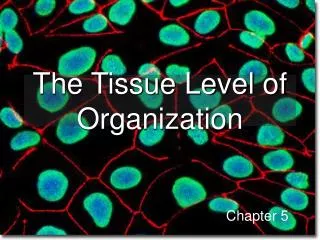
The Tissue Level of Organization Tissues Dr. Nabil Khouri
Learning Objectives • Identify the four major tissue types and describe their functions. • Describe the relationship between form and function for each tissue type. • Discuss the types and functions of epithelial tissues.
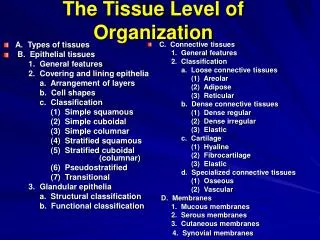
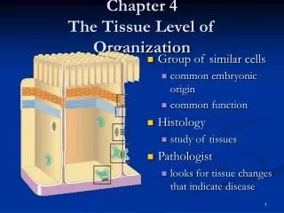
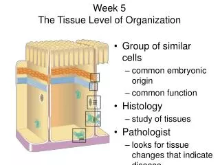
Tissues of the Body: An Introduction Tissues and tissue types • Tissues are: • Collections of specialized cells and cell products organized to perform a limited number of functions • The four tissue types are: • Epithelial • Connective • Muscular • Nervous
Embryonic Tissues Embryo begins as a single cell divides into many cells that form layers (strata) Three primary germ layers Ectoderm (outer) gives rise to: epidermis & nervous system Endoderm (inner): mucous membranes: GI tract and respiratory linings; digestive glands. Mesoderm (middle) forms mesenchyme (gelatinuous tissue) and then give rise to muscle, bone, and blood
Tissues = groups of cells that are similar in structure and function. • Epithelium • Coverings • Linings of surfaces • Connective • Support • Bone, ligaments, fat • Muscle • Movement • Nervous • Control • Brain, nerves, spinal cord
Function of Epithelial Tissue • Protection • Skin protects from sunlight & bacteria & physical damage. • Absorption • Lining of small intestine, absorbing nutrients into blood • Filtration • Lining of Kidney tubules filtering wastes from blood plasma • Secretion • Different glands produce perspiration, oil, digestive enzymes and mucus
Characteristics of Epithelial Tissue • Form continuous sheets (fit like tiles) • Apical Surface • All epithelial cells have a top surface that borders an open space – known as a lumen • Basement Membrane • Underside of all epithelial cells which anchors them to connective tissue • Avascularity (a = without) • Lacks blood vessels • Nourished by connective tissue • Regenerate & repair quickly
Epithelial tissue • Includes epithelium & glands • Glands are secretory epithelium lining organs • The epithelium : • Is avascular • Forms a protective barrier that regulates permeability • Cells may show polarity
Maintaining the integrity of epithelium • Cells attach via cell adhesion molecules (CAM) • Cells are attach at specialized cell junctions • Tight junctions • Desmosomes • Gap junctions
Structure of typical epithelium • Basal lamina attaches to underlying surface • Lamina lucida • Lamina densa • Germinative cells replace short-lived epithelial cells
Classification of epithelia • Cell Shape • Squamous – flattened like fish scales • Cuboidal - cubes • Columnar - columns • Cell Layers (NUMBER) • Simple (one layer) • Stratified (many layers) • It is then named for the type of cell at the apical surface.
Simple Squamous Epithelium • Mesothelia or Endothelia • Structure • Single Layer of flattened cells • Function • Absorption, secretion permeability and filtration • Reduce frection • Not effective protection – single layer of cells. • Location • Walls of capillaries, air sacs in lungs • Form serous membranes in body cavity
Stratified Squamous Epithelium • Structure • Many layers (usually cubodial/columnar at bottom and squamous at top) • Function • Physical Protection • Keratin (protein) is accumulated in older cells near the surface – waterproofs and toughens skin. • Location • Skin (keratinized), mouth & throat
Simple Cuboidal Epithelium • Structure • Single layer of cube shaped cells • Function • Secretion and transportation in glands, filtration in kidneys • Location • Glands and ducts (pancreas & salivary), kidney tubules, covers ovaries
Stratified Cuboidal Epithelia Stratified Cubodial (layers of cubodial only)
Simple Columnar Epithelium • Structure • Elongated layer of cells with nuclei at same level • Function • Absorption, Protection & Secretion • When open to body cavities called mucous membranes • Special Features • Microvilli, bumpy extension of apical surface, increase surface area and absorption rate. • Goblet cells, single cell glands, produce protective mucus. • Location • Linings of entire digestive tract
Stratified columnar epithelium • Is a rare type of epithelial tissue composed of column shaped cells arranged in multiple layers. • Stratified columnar epithelia are found in the ocular conjunctiva of the eye, in parts of the pharynx and anus, the female's uterus, the male urethra and vas deferens. Also found in Lobar ducts in salivary glands. • The cells function in secretion and protection.
Pseudostratified Epithelium • Structure • Irregularly shaped cells with nuclei at different levels – appear stratified, but It is not. • All cells reach basement membrane • Function • Absorption and Secretion • Goblet cells produce mucus • Cilia (larger than microvilli) sweep mucus • Location • Respiratory Linings & Reproductive tract
Transitional Epithelium • Structure • Many layers • Very specialized – cells at base are cuboidal or columnar, at surface will vary. • Change between stratified & simple as tissue is stretched out. • Function • Allows stretching (change size) • Location • Urinary bladder, ureters & urethra
Glands • Unicellular • Individual secretory cells • Multicellular • Organs containing glandular epithelium • Classified according to structure
Glands • One or more cells that make and secrete a product. • Secretion = protein in aqueous solution: hormones, acids, oils. • Endocrine glands • No duct, release secretion into blood vessels • Often hormones • Thyroid, adrenal and pituitary glands • Exocrine glands • Contain ducts, empty onto epithelial surface • Sweat, Oil glands, Salivary glands, Mammary glands.
a. Sweet Glands There are 2 types of sweat glands: • Eccrine sweat glands, all over body except lips and part of external genitalia; • Apocrine sweat glands, only in axilla, areola, nipple of mammary gland, and circumanal region and the external genitalia. The ceruminous glands of ear and glands of Moll of eyelid are also apocrine. • Both the Eccrine and the Apocrine sweat glands are innervated by the sympathetic nervous system. • Eccrine glands respond differently to heat and nervous state. • The apocrine glands respond to emotional and sensory stimuli but not heat.
Eccrine Sweat Glands • Are simple coiled glands that regulate body temperature. • The secretory segment is deep in the dermis or upper hypodermis. • Its duct leads to surface. • In the secretory region there are clear cells that produce the watery component of sweat and dark cells that produce a protein secretion. • Duct cells form the walls from the secretory portion to the area near the surface where the epidermal cells form the wall. • The duct is stratified cuboidal epithelium .
b. Sebaceous Glands or Oil GlandsFound in the skin of mammals and these glands secrete sebum. Sebum is made of fat (lipids) and the debris of dead fat-producing cells. • These glands exist in humans throughout the skin except in the palms of the hands and soles of the feet. • Sebum acts to protect and waterproof hair and skin, and keep them from becoming dry, brittle, and cracked. It can also inhibit the growth of microorganisms on skin.
c. Ceruminous glands • Are involved in skin problems such as acne and keratosis pilaris. A blocked sebaceous gland can result in a sebaceous cyst. • Earwax, also known by the medical term cerumen, is a yellowish, waxy substance secreted in the ear canal of humans and many other mammals. It plays a vital role in the human ear canal, assisting in cleaning and lubrication, and also provides some protection from bacteria, fungus, and insects
D. Mammary glands Are the organs that, in the female mammal, produce milk for the sustenance of the young. These Exocrine glands are enlarged and modified sweat glands and are the characteristic of mammals which gave the class its name.
The human mammary glands are modified sweat glands and are developed from two sources --the parenchyma (alveoli and ductules ) from the surface ectoderm ,the fibrofatty stroma from the underlying endoderm. • At birth mammary glands of both sexes remain in infantile form .This condition persists throughout life in normal male. • First change is seen at puberty in females ,in the form of deposition of fat and increase in size and attain hemispherical outlines. With the start of reproductive cycle after puberty ,glandular tissue show changes with the alteration in concentration of oestrogen and progesterone in each cycle. • Ultimately during pregnancy final maturation of the glands takes place and they are ready for milk secretion under the influence of oestrogen ,progesterone ,prolactin and probably hCG.. • Some milk is secreted into the ducts as early as 5 months but the amount is less compared to large amount secreted at child birth.Milk is secreted within an hour of child birth and first formed milk is called colostrum which is yellowish in colour and rich in protein and antibodies and provide immunity to the baby.Normal milk production starts 2-3 days after child birth.
Glandular secretions can be: • Merocrine (product released through exocytosis) • Apocrine (involves the loss of both product and cytoplasm) • Holocrine (destroys the cell)
Apocrine Sweat Glands • The secretory product from the gland. • The duct has a narrow lumen. • Apocrine secretions contain protein, carbohydrate, ammonia and lipid.
Merocrine gland • Cells that secrete products via the merocrine method form membrane-bound secretory vesicles internal to the cell. • These are moved to the apical surface where the vesicles coalesce with the membrane on the apical surface to release the product. Most glands release their products in way.
Holocrine Glands • The third type of secretory release, Holocrine, involves death of the cell. The secretory cell is released and as it breaks apart, the contents of the cell become the secretory product. This mode of secretion results in the most complex secretory product. Some sweat glands located in the axillae, pubic areas, and around the areoli of the breasts release their products in this manner. Sebaceous glands also are of this type.