Download
1 / 127
1.31k likes | 1.83k Vues
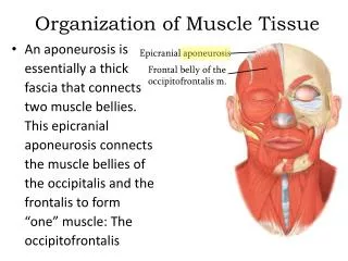
4 Tissue Level of Organization. Section 1: Epithelial Tissue. Learning Outcomes 4.1 Describe epithelial tissues, including cell shape, layers, and functions. 4.2 Discuss the types and functions of intercellular connections between epithelial cells.

E N D
4 Tissue Level of Organization
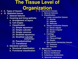
Section 1: Epithelial Tissue Learning Outcomes 4.1 Describe epithelial tissues, including cell shape, layers, and functions. 4.2 Discuss the types and functions of intercellular connections between epithelial cells. 4.3 Describe the structure and function of squamous epithelium. 4.4 Describe the structure, function, and locations of cuboidal and transitional epithelia.
Learning Outcomes 4.5 Describe the structure, function, and locations of columnar epithelia. 4.6 Describe the structure, function, and locations of glandular epithelia. Section 1: Epithelial Tissue
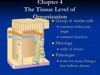
Atoms Molecules Cells Tissues Chemical level imaged only with special techniques Cellular level imaged often with electron microscope Body contains trillions of cells Only ~200 types of cells Tissue level can be imaged with light microscope Section 1: Epithelial Tissue
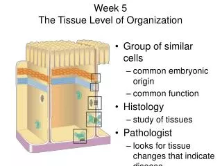
Tissues (cells working together) Histology (study of tissues) Four basic types Epithelial Connective Muscle Neural Section 1: Epithelial Tissue
Figure 4 Section 1 The tissue level of organization, which consists of four tissue types The Chemical Level combine to form MOLECULES ATOMS interact to form The Cellular Level that secrete and regulate EXTRACELLULAR MATERIAL AND FLUIDS CELLS combine to form The Tissue Level TISSUES with special functions can be classified as MUSCLE TISSUE EPITHELIAL TISSUE NEURAL TISSUE CONNECTIVE TISSUE • Covers exposed surfaces • Contracts to produce active movement • Conducts electrical impulses • Fills internal spaces • Lines internal passageways and chambers • Provides structural support • Carries information • Produces glandular secretions • Stores energy
Module 4.1: Epithelial tissue Epithelial tissue Epithelia Cover exposed surfaces and internal cavities/passageways Often contain secretory or gland cells Scattered among other cell types Glands (derived from epithelia but more secretory cells) Two types Exocrine glands Secrete on external areas Endocrine glands Secrete hormones into interstitial fluid
Figure 4.1 1 The components of epithelial tissue Epithelial Tissue Includes Epithelia Glands Epithelia cover exposed surfaces and line internal cavities and passageways; they often contain secretory cells, or gland cells, scattered among the other cell types. Glands are derived from epithelia, but secretory cells predominate; there are two types: Exocrine Glands Endocrine Glands Exocrine glands secrete onto external surfaces or into internal passageways (ducts) that connect to the exterior. Endocrine glands secrete hormones or precursors into the interstitial fluid, usually for distribution by the bloodstream.
Functions of epithelial tissue Provide physical protection Control permeability Provide sensation Produce specialized secretions Module 4.1: Epithelial tissue
Basic features of epithelial cells Apical surface (faces exterior or internal space) Microvilli often found on cells of digestive, urinary, and reproductive tracts Cilia often found on cells lining respiratory and some parts of reproductive tracts Faces lumen (space) when lining hollow organs Base (attached to adjacent tissues) Basolateral surfaces Includes base and lateral surfaces (attached to neighboring cells) Have membranous organelles comparable to other cell types Module 4.1: Epithelial tissue
Three epithelial cell shapes (perpendicular section) Squamous (thin and flat) Cuboidal (small boxes) Columnar (slender rectangles) Can be layered Single layer (simple epithelium) Several layers (stratified epithelium) Generally located in areas that need protection Module 4.1: Epithelial tissue
Module 4.1 Review a. List four essential functions of epithelial tissue. b. Summarize the classification of an epithelium based on cell shape and number of cell layers. c. What is the probable function of an epithelial surface whose cells bear many cilia?
Module 4.2: Epithelial cells are extensively interconnected Many types of connections to form complete cover or lining Have ability to replace damaged or lost cells Lack blood vessels (avascular) Lowest cell layers must remain attached to underlying tissues to be near blood vessels Animation: Intercellular Connections
Figure 4.2 1 Microvilli The structures that connect epithelial cells to each other and to adjacent tissues APICAL SURFACE Intercellular attachments Occluding junctions form a barrier that isolates the basolateral surfaces and deeper tissues from the contents of the lumen. An adhesion belt locks together the terminal webs of neighboring cells, strengthening the apical region and preventing distortion and leakage at the occluding junctions. Gap junctions permit chemical communication that coordinates the activities of adjacent cells. Desmosomes (DEZ-mō-sōms; desmos, ligament + soma, body) provide firm attachment between neighboring cells by interlocking their cytoskeletons. BASE Basal Lamina Hemidesmosome
Hemidesmosomes Made of peripheral and transmembrane proteins Attach deepest epithelial cells to basal lamina Basal lamina (basement membrane) layers Clear layer (lamina lucida) Contains glycoproteins and fine protein filaments Dense layer (lamina densa) Contains bundles of coarse protein fibers Gives strength and restricts diffusion Module 4.2: Epithelial cells are extensively interconnected
Figure 4.2 2 The structures that connect epithelial cells to each other and to adjacent tissues Basal Lamina Hemidesmosome Intermediate filaments of the cytoskeleton The basal lamina, or basement membrane, is a complex structure produced by the basal surface of the epithelium and the underlying connective tissue. The clear layer, or lamina lucida (LAM-i-nah LOO-si-dah; lamina, thin layer + lucida, clear) contains glycoproteins and a network of fine protein filaments. The dense layer, or lamina densa, containing bundles of coarse protein fibers, gives the basal lamina its strength and acts as a filter that restricts diffusion between the adjacent tissues and the epithelium. A hemidesmosome, which attaches the deepest epithelial cells to the basal lamina
Types of intercellular attachments Occluding junctions Form barrier that prevents lumen contents from getting past cells Adjacent plasma membranes tightly bound with proteins Adhesion belt Attaches terminal webs of adjacent cells Reinforces occluding junctions Dense protein band surrounding cell Module 4.2: Epithelial cells are extensively interconnected
Figure 4.2 3 The structures that connect epithelial cells to each other and to adjacent tissues At an occluding junction, the lipid portions of the two plasma membranes are tightly bound together by interlocking membrane proteins. An occluding junction
Figure 4.2 4 The structures that connect epithelial cells to each other and to adjacent tissues An adhesion belt
Types of intercellular attachments (continued) Gap junctions Permit chemical communication to coordinate activities of adjacent cells Formed by interlocking junctional proteins (connexons) Examples: ciliated epithelial tissues, cardiac muscle tissue Desmosomes (desmos, ligament + soma, body) Interlock cytoskeletons of adjacent cells Very strong Formed by: Cell adhesion molecules (CAMs) Intercellular cement (thin layer of proteoglycans, notably hyaluronan) Module 4.2: Epithelial cells are extensively interconnected
Figure 4.2 5 The structures that connect epithelial cells to each other and to adjacent tissues Connexons are channel proteins that form a narrow passageway and let small molecules and ions pass from cell to cell. An jap junction
Figure 4.2 6 The structures that connect epithelial cells to each other and to adjacent tissues Cell adhesion molecules (CAMs) are transmembrane proteins that bind to each other and to extracellular materials. The membranes of adjacent cells may also be bonded by intercellular cement, a thin layer of proteoglycans that contain polysaccharide derivatives, most notably hyaluronan. A desmosome
Module 4.2 Review a. Identify the various types of epithelial intercellularconnections. b. How do epithelial tissues, which are avascular, obtain needed nutrients? c. What is the functional significance of gap junctions?
Module 4.3: Squamous epithelia Simple squamous epithelium (squama, plate or scale) Thin and flat, irregular in shape Surface view: like fried eggs side to side Sectional view: jigsaw pieces with disc-shaped nucleus Found in protected regions Where absorption or diffusion takes place Examples: along kidney passages, inside eye, alveoli of the lung Where a slippery surface reduces friction Lining ventral body cavity (mesothelium) Lining heart and blood vessels (endothelium)
Figure 4.3 1 Simple squamous epithelia, which include the specially named endothelium and mesothelium Endothelium lining the inside of the heart and blood vessels Mesothelium lining the pericardial cavity and the peritoneal cavity Connective tissue Nucleus Cytoplasm Sectioned epithelial cell The surface of a simple squamous epithelium Section of peritoneum Simple squamous epithelium LM x 270
Module 4.3: Squamous epithelia Stratified squamous epithelium Located where mechanical or chemical stresses are severe Series of layers Example locations: skin, mouth, throat, esophagus, rectum, anus, vagina
Figure 4.3 2 A stratified squamous epithelium Squamous superficial cells Stem cells Basal lamina Connective tissue Surface of the tongue LM x 400
Stratified squamous epithelium Two types Keratinized (with keratin proteins) Tough and water resistant Found at surface of skin Nonkeratinized Resists abrasion but can dry out Found in oral cavity, pharynx, esophagus, anus, vagina Module 4.3: Squamous epithelia
Figure 4.3 3 The structure of a keratinized stratified squamous epithelium Keratinized skin cells Keratin fibers Surface of human skin
Module 4.3 Review a.What properties are common to keratinized epithelia? b. Why do the pharynx, esophagus, anus, and vagina have a similar epithelial organization? c. Under a light microscope, simple squamous epithelium is seen on the outer surface. Could this be a skin surface sample? Why or why not?
Module 4.4: Cuboidal and transitional epithelia Cuboidal epithelium Cells resemble hexagonal boxes with nucleus in center Types Simple cuboidal epithelium Locations Lining exocrine glands and ducts Portions of kidney Secretory chambers of thyroid gland Stratified cuboidal epithelium Rare Locations Ducts of various exocrine glands
Figure 4.4 1 – 3 Examples of cuboidal epithelia Connective tissue Basal lamina Simple cuboidal cells Lumen of duct Nucleus LM x 1400 A cuboidal epithelium The simple cuboidal epithelium in a sectioned kidney tubule Lumen of duct Stratified cuboidal cells Basal lamina Nuclei Connective tissue LM x 1413 The stratified cuboidal epithelium in a sweat gland duct
Transitional epithelium Unusual stratified epithelium that stretches and recoils Transitional = changes Locations Urinary bladder Urethra Urine-collecting chambers of kidneys Module 4.4: Cuboidal and transitional epithelia
Figure 4.4 4 The transitional epithelium in an empty and a full urinary bladder Epithelium in a Relaxed Bladder In an empty urinary bladder, the superficial cells are cuboidal with a dome-shaped surface. Epithelium (relaxed) Basal lamina LM x 400 Connective tissue and smooth muscle layers Relaxed bladder
Module 4.4 Review a.Identify the epithelium that lines the urinary bladder and changes in appearance as stretching occurs. b. Describe the appearance of simple cuboidal epithelial cells in sectional view. c. Stratified cuboidal epithelia are associated with what epithelial structures?
Module 4.5: Columnar epithelia Columnar epithelium Cells appear rectangular in sectional view Elongated nuclei near basal lamina Types Simple columnar epithelium Locations Stomach Intestine Uterine tubes Kidney ducts
Figure 4.5 1 The simpler columnar epithelium in the intestinal lining Microvilli Cytoplasm Nucleus Basal lamina Loose connective tissue LM x 350
Columnar epithelium Types (continued) Pseudostratified columnar epithelium Varying cell shapes and functions Nuclei located at different areas of cell, so appears stratified Every cell attached to basal lamina Cells typically possess cilia Locations Nasal cavities Trachea Larger airways of lungs Module 4.5: Columnar epithelia
Figure 4.5 2 The pseudostratified columnar epithelium in the trachea Cilia Cytoplasm Nuclei Basal lamina Loose connective tissue LM x 394
Columnar epithelium Types (continued) Stratified columnar epithelium Relatively rare Either two or multiple layers In multiple layers, only superficial cells are columns Located in large ducts of salivary glands and pancreas Module 4.5: Columnar epithelia
Figure 4.5 3 The stratified columnar epithelium in a salivary gland duct Loose connective tissue Basal cells Superficial columnar cells Lumen Lumen Cytoplasm Nuclei Basal lamina LM x 175
Module 4.5 Review a. Describe the appearance of simple columnarepithelial cells in a sectional view. b. Explain why a pseudostratified columnar epithelium is not truly stratified. c. The columnar epithelium lining the intestine typically has ___________ on its apical surface.
Module 4.6: Glandular epithelia Glands Collections of epithelial cells that produce secretions Can be scattered cells or complex organs Two types of glandular organs Endocrine glands (secretions into interstitial fluid) More information in later chapter Exocrine glands (secretions into ducts that open onto epithelial surface)
Exocrine glands Three types of secretion Merocrine secretion (meros, part) Product released from secretory vesicles by exocytosis Most common type of secretion Mucin Merocrine secretion that mixes with water to form mucus Used as lubricant, protective barrier, and trap for foreign particles and microorganisms Module 4.6: Glandular epithelia
Exocrine glands Three types of secretion (continued) Apocrine secretion (apo-, off) Loss of apical surface and cytoplasm with secretion Example: mammary glands Merocrine and apocrine secretions in milk Holocrine secretion (holos, entire) Entire cell bursts, releasing secretion and killing cell Replaced by stem cell division Module 4.6: Glandular epithelia Animation: Mechanisms of Glandular Secretion
Figure 4.6 1 Mucin is a merocrine secretion that mixes with water to form mucus. Mucus is an effective lubricant, a protective barrier, and a sticky trap for foreign particles and microorganisms. Secretory vesicle (containing mucin) Golgi apparatus Nucleus TEM x 3120 The three types of secretion by glandular epithelial cells In merocrine secretion (MER-u-krin; meros, part), the product is released from secretory vesicles by exocytosis. This is the most common mode of secretion. Breaks down Regrowth Salivary gland Secretion Golgi apparatus Mammary gland Apocrine secretion (AP-ō-krin; apo-, off) involves the loss of cytoplasm as well as the secretory product. The apical portion of the cytoplasm becomes packed with secretory vesicles and is then shed. Milk production in the mammary glands involves a combination of merocrine and apocrine secretions. Cells burst, releasing cytoplasmic contents. Hair Cells produce secretion, increasing in size. Sebaceous gland Hair follicle Start Cell division replaces lost cells. Stem cell Holocrine secretion (HOL-ō-krin; holos, entire), by contrast, destroys the gland cell. During holocrine secretion, the entire cell becomes packed with secretory products and then bursts, releasing the secretion and killing the cell. Further secretion depends on the replacement of destroyed gland cells by the division of stem cells.
Multicellular exocrine gland structure Gland classification Based on duct structure Simple (single duct that does not divide) Compound (duct divides one or more times) Based on structure of secretory area Tubular (glandular cells form tubes) Alveolar or acinar (glandular cells form sacs) Tubuloalveolar (glandular cells form tubes and sacs) Module 4.6: Glandular epithelia
Figure 4.6 2 A gland is branched if several secretory areas (tubular or acinar) share a duct. Note that “branched” refers to the glandular areas and not to the duct. Simple exocrine glands Duct Gland cells SIMPLE TUBULAR SIMPLE ALVEOLAR (ACINAR) SIMPLE BRANCHED ALVEOLAR SIMPLE BRANCHED TUBULAR SIMPLE COILED TUBULAR Examples: Examples: Examples: Examples: Examples: • A stage in the em- bryonic development of simple branched glands • Sebaceous (oil) glands • Intestinal glands • Merocrine sweat glands • Gastric glands • Mucous glands of esophagus, tongue, duodenum Glands whose glandular cells form tubes are tubular; the tubes may be straight or coiled. Glands whose glandular cells form sac-like pockets are alveolar (al-VĒ-ō-lar; alveolus, sac) or acinar (AS-i-nar; acinus, chamber).
Figure 4.6 3 Compound exocrine glands Glands whose secretory cells form both tubes and sacs are called tubuloalveolar. COMPOUND TUBULAR COMPOUND ALVEOLAR (ACINAR) COMPOUND TUBULOALVEOLAR Examples: Examples: Examples: • Mammary glands • Salivary glands • Mucous glands (in mouth) • Glands of respiratory passages • Bulbo-urethral glands (in male reproductive system) • Pancreas • Seminiferous tubules of testes
Unicellular glands Individual, scattered secretory cells (mucous cells) Secrete mucin Apical cytoplasm filled with large secretory vesicles Module 4.6: Glandular epithelia