Ionization and Mass Analyzers
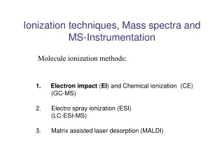
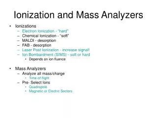
Ionization and Mass Analyzers. Ionizations Electron Ionization - “hard” Chemical Ionization - “soft” MALDI - desorption FAB - desorption Laser Post Ionization - increase signal! Ion Bombardment (SIMS) - soft or hard Depends on ion fluence Mass Analyzers Analyze all mass/charge

Ionization and Mass Analyzers
E N D
Presentation Transcript
Ionization and Mass Analyzers • Ionizations • Electron Ionization - “hard” • Chemical Ionization - “soft” • MALDI - desorption • FAB - desorption • Laser Post Ionization - increase signal! • Ion Bombardment (SIMS) - soft or hard • Depends on ion fluence • Mass Analyzers • Analyze all mass/charge • Time-of-flight • Pre- Select Ions • Quadropole • Magnetic or Electric Sectors
Electron Ionization • Molecule + e- molecule+ + 2e- • Four electromagnetic poles • Rapidly oscillating field • If mass of ion and frequency of oscillation are comparable then the ion will oscillate towards the detector and be measured • A certain mass range can be selected depending on the pole diameter Quadropole Mass Analyzer
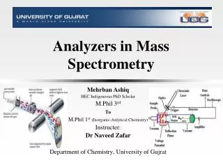
mass separate focus Electrostatic and Magnetic Sectors B = strength of magnetic field V = ion accelerating voltage r = radius of ion curvature m = mass of ion z = charge of ion M = 1-50 (change by changing B) http://www.cea.com/cai/simsinst/m_anal.htm
20 particles 10 ion - + e- Secondary Ion Mass Spectrometry “Ionization” Ga+ Static SIMS = < 1 x 10¹³ ions/cm2
Time-of-Flight Analyzer d -V +V M+ MCP detector Field Free Region Sample e = charge on an electron 1.6 x 10⁻¹⁹ C t = time for ion to reach detector m = mass v = velocity V = stage voltage d = length of field free region V = voltage of detector
Laser Postionization (LPI) Laser Beam Postionization parameters: 800 nm, 150 fs laser pulses, 8.5 x 10¹² W/cm2 power density
TOF-SIMS Molecule-Specific Imaging total ion image Ag+ Intensity Ion Gun y x 570 µm x 570 µm m/z, +ions molecule-specific image Si+ Ag+ Cu+ Si+ Cu+ copper grid affixed to silicon substrate with silver paste
TOF-SIMS Identification of Plasma Membrane Lipids * - * +2H m/z 552 m/z 142 + cholesterol m/z 369 m/z 385 (M-H)+ DPPE – dipalmitoylphosphatidylethanolamine m/z 682 (M+H)+
Are there Lipid Domains in the Inner Leaflet of the Membrane? cholesterol DPPE Cholesterol DPPE + 552 Total Ion DPPE/Cholesterol 250 µm x 250 µm Intensity Au - A. G. Sostarecz; C. M. McQuaw et. al. J. Am. Chem. Soc.2004. C. M. McQuaw; A. G. Sostarecz et. al. Langmuir2005.
Multi-Isotope Imaging Mass Spectrometry Atomic Mass Images 14N nucleus lamellipodia endothelial cell Image from Claude Lechene Harvard Medical School
mass separate focus 20 particles 10 ion - + e- MIMS is a Secondary Ion Mass Spectrometry Technique B = strength of magnetic field V = ion accelerating voltage r = radius of ion curvature m = mass of ion z = charge of ion M = 1-50 (change by changing B) http://www.cea.com/cai/simsinst/m_anal.htm
MIMS uses an Electrostatic and Magnetic Sector Mass Analyzer mass separate focus 14N ion 1 84 % 16 % Image from Claude Lechene *** MIMS analysis is performed as a collaborative project with Dr. Claude Lechene at the National Resource for Imaging Mass Spectrometry at Harvard Medical School