Microscopy and Cell Structure
Microscopy and Cell Structure. Chapter 3. Microscope Techniques Microscopes. Microscopes Most important tool for studying microorganisms Use viable light to observe objects Magnify images approximately 1,000x Electron microscope, introduced in 1931, can magnify images in excess of 100,000x

Microscopy and Cell Structure
E N D
Presentation Transcript
Microscopy and Cell Structure Chapter 3
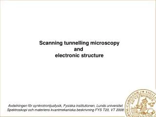
Microscope TechniquesMicroscopes • Microscopes • Most important tool for studying microorganisms • Use viable light to observe objects • Magnify images approximately 1,000x • Electron microscope, introduced in 1931, can magnify images in excess of 100,000x • Scanning probe microscope, introduced in 1981, can view individual atoms
Principles of Light Microscopy • Light Microscopy • Light passes through specimen, then through series of magnifying lenses • Most common and easiest to use is the bright-field microscope • Important factors in light microscopy include • Magnification • Resolution • Contrast
Principles of Light Microscopy • Magnification • Microscope has two magnifying lenses • Called compound microscope • Lens include • Ocular lens and objective lens • Most bright field scopes have four magnifications of objective lenses, 4x, 10x, 40x and 100x • Lenses combine to enlarge objects • Magnification is equal to the factor of the ocular x the objective • 10x X 100x = 1,000x
Principles of Light Microscopy • Magnification • Bright field scopes have condenser lens • Has no affect on magnification • Used to focus illumination on specimen
Principles of Light Microscopy • Resolution • Usefulness of microscope depends on its ability to resolve two objects that are very close together • Resolving power is defined as the minimum distance existing between two objects where those objects still appear as separate objects • Resolving power determines how much detail can be seen
Principles of Light Microscopy • Resolution • Resolution depends on the quality of lenses and wavelength of illuminating light • How much light is released from the lens • Maximum resolving power of most brightfield microscopes is 0.2 μm (1x10⁻⁶) • This is sufficient to see most bacterial structures • Too low to see viruses
Principles of Light Microscopy • Resolution • Resolution is enhanced with lenses of higher magnification (100x) by the use of immersion oil • Oil reduces light refraction • Light bends as it moves from glass to air • Oil bridges the gap between the specimen slide and lens and reduces refraction • Immersion oil has nearly same refractive index as glass
Principles of Light Microscopy • Contrast • Reflects the number of visible shades in a specimen • Higher contrast achieved for microscopy through specimen staining
Principles of Light Microscopy • Examples of light microscopes that increase contrast • Phase-Contrast Microscope • Interference Microscope • Dark-Field Microscope • Fluorescence Microscope • Confocal Scanning Laser Microscope
Principles of Light Microscopy • Phase-Contrast • Amplifies differences between refractive indexes of cells and surrounding medium • Uses set of rings and diaphragms to achieve resolution
Principles of Light Microscopy • Interference Scope • This microscope causes specimen to appear three dimensional • Depends on differences in refractive index • Most frequently used interference scope is Nomarski differential interference contrast
Principles of Light Microscopy • Dark-Field Microscope • Reverse image • Specimen appears bright on a dark background • Like a photographic negative • Achieves image through a modified condenser
Principles of Light Microscopy • Fluorescence Microscope • Used to observe organisms that are naturally fluorescent or are flagged with fluorescent dye • Fluorescent molecule absorbs ultraviolet light and emits visible light • Image fluoresces on dark background
Principles of Light Microscopy • Confocal Scanning Laser Microscope • Used to construct three dimensional image of thicker structures • Provides detailed sectional views of internal structures of an intact organism • Laser sends beam through sections of organism • Computer constructs 3-D image from sections
Principles of Light Microscopy • Electron Microscope • Uses electromagnetic lenses, electrons and fluorescent screen to produce image • Resolution increased 1,000 fold over brightfield microscope • To about 0.3 nm (1x10⁻⁹) • Magnification increased to 100,000x • Two types of electron microscopes • Transmission • Scanning
Principles of Light Microscopy • Transmission Electron Microscope (TEM) • Used to observe fine detail • Directs beam of electrons at specimen • Electrons pass through or scatter at surface • Shows dark and light areas • Darker areas more dense • Specimen preparation through • Thin sectioning • Freeze fracturing or freeze etching
Principles of Light Microscopy • Scanning Electron Microscope (SEM) • Used to observe surface detail • Beam of electrons scan surface of specimen • Specimen coated with metal • Usually gold • Electrons are released and reflected into viewing chamber • Some atomic microscopes capable of seeing single atoms
Microscope TechniquesDyes and Staining • Dyes and Staining • Cells are frequently stained to observe organisms • Satins are made of organic salts • Dyes carry (+) or (-) charge on the molecule • Molecule binds to certain cell structures • Dyes divided into basic or acidic based on charge • Basic dyes carry positive charge and bond to cell structures that carry negative charge • Commonly stain the cell • Acidic dyes carry positive charge and are repelled by cell structures that carry negative charge • Commonly stain the background
Microscope TechniquesDyes and Staining • Basic dyes (+) more commonly used than acidic dyes (-) • Common basic (+) dyes include • Methylene blue • Crystal violet • Safrinin • Malachite green
Microscope TechniquesDyes and Staining • Staining Procedures • Simple stain uses one basic stain to stain the cell • Allows for increased contrast between cell and background • All cells stained the same color • No differentiation between cell types
Microscope TechniquesDyes and Staining • Differential Stains • Used to distinguish one bacterial group from another • Uses a series of reagents • Two most common differential stains • Gram stain • Acid-fast stain
Microscope TechniquesDyes and Staining • Gram Stain • Most widely used procedure for staining bacteria • Developed over century ago • Dr. Hans Christian Gram • Bacteria separated into two major groups • Gram positive • Stained purple • Gram negative • Stained red or pink
Dyes and Staining • The Gram Stain
Microscope TechniquesDyes and Staining • Acid-fast Stain • Used to stain organisms that resist conventional staining • Used to stain members of genus Mycobacterium • High lipid concentration in cell wall prevents uptake of dye • Uses heat to facilitate staining • Once stained difficult to decolorize
Microscope TechniquesDyes and Staining • Acid-fast Stain • Can be used for presumptive identification in diagnosis of clinical specimens • Requires multiple steps • Primary dye • Carbol fuchsin • Colors acid-fast bacteria red • Decolorizer • Generally acid alcohol • Removes stains from non acid-fast bacteria • Counter stain • Methylene blue • Colors non acid-fast bacteria blue
Microscope TechniquesDyes and Staining • Special Stains • Capsule stain • Example of negative stain • Allows capsule to stand out around organism • Endospore stain • Staining enhances endospore • Uses heat to facilitate staining • Flagella stain • Staining increases diameter of flagella • Makes more visible
Morphology of Prokaryotic Cells • Prokaryotes exhibit a variety of shapes • Most common • Coccus • Spherical • Bacillus • Rod or cylinder shaped • Cell shape not to be confused with Bacillus genus
Morphology of Prokaryotic Cells • Prokaryotes exhibit a variety of shapes • Other shapes • Coccobacillus • Short round rod • Vibrio • Curved rod • Spirillum • Spiral shaped • Spirochete • Helical shape • Pleomorphic • Bacteria able to vary shape
Morphology of Prokaryotic Cells • Prokaryotic cells may form groupings after cell division • Cells adhere together after cell division for characteristic arrangements • Arrangement depends on plan of division • Especially in the cocci
Morphology of Prokaryotic Cells • Division along a single plane may result in pairs or chains of cells • Pairs = diplococci • Example: Neisseria gonorrhoeae • Chains = streptococci • Example: species of Streptococcus
Morphology of Prokaryotic Cells • Division along two or three perpendicular planes form cubical packets • Example: Sarcina genus • Division along several random planes form clusters • Example: species of Staphylococcus
Morphology of Prokaryotic Cells • Some bacteria live in groups with other bacterial cells • They form multicellular associations • Example: myxobacteria • These organisms form a swarm of cells • Allows for the release of enzymes which degrade organic material • In the absence of water cells for fruiting bodies • Other organisms for biofilms • Formation allows for changes in cellular activity
Cytoplasmic Membrane • Cytoplasmic membrane • Delicate thin fluid structure • Surrounds cytoplasm of cell • Defines boundary • Serves as a semi permeable barrier • Barrier between cell and external environment
Cytoplasmic Membrane • Structure is a lipid bilayer with embedded proteins • Bilayer consists of two opposing leaflets • Leaflets composed of phospholipids • Each contains a hydrophilic phosphate head and hydrophobic fatty acid tail
The Basic Structural Component of the Membrane: Phospholipid Molecule
Cytoplasmic Membrane • Membrane is embedded with numerous protein • More that 200 different proteins • Proteins function as receptors and transport gates • Provides mechanism to sense surroundings • Proteins are not stationary • Constantly changing position • Called fluid mosaic model
Cytoplasmic Membrane • Cytoplasmic membrane is selectively permeable • Determines which molecules pass into or out of cell • Few molecules pass through freely • Molecules pass through membrane via simple diffusion or transport mechanisms that may require carrier proteins and energy
Cytoplasmic Membrane • Simple diffusion • Process by which molecules move freely across the cytoplasmic membrane • Water, certain gases and small hydrophobic molecules pass through via simple diffusion
Cytoplasmic Membrane • Simple diffusion • Osmosis • The ability of water to flow freely across the cytoplasmic membrane • Water flows to equalize solute concentrations inside and outside the cell • Inflow of water exerts osmotic pressure on membrane • Membrane rupture is prevented by rigid cell wall of bacteria
Cytoplasmic Membrane • Membrane also the site of energy production • Energy produced through series of embedded proteins • Electron transport chain • Proteins are used in the formation of proton motive force • Energy produced in proton motive force is used to drive other transport mechanisms
Cytoplasmic Membrane • Directed movement across the membrane • Movement of many molecules directed by transport systems • Transport systems employ highly selective proteins • Transport proteins (a.k.a permeases or carriers) • These proteins span membrane • Single carrier transports specific type molecule • Most transport proteins are produced in response to need • Transport systems include • Facilitated diffusion • Active transport • Group translocation
Cytoplasmic Membrane • Facilitated diffusion • Moves compounds across membrane exploiting a concentration gradient • Flow from area of greater concentration to area of lesser concentration • Molecules are transported until equilibrium is reached • System can only eliminate concentration gradient it cannot create one • No energy is required for facilitated diffusion • Example: movement of glycerol into the cell
Cytoplasmic Membrane • Active transport • Moves compounds against a concentration gradient • Requires an expenditure of energy • Two primary mechanisms • Proton motive force • ATP Binding Cassette system