Download
1 / 18
250 likes | 825 Vues
MUSCLES INVOLVED IN RESPIRATION. Dr. Ahmed Fathalla Ibrahim & Dr. Zeenat Zaidi. OBJECTIVES. At the end of the lecture, students should be able to: Describe the components of the thoracic cage and their articulations. Describe in brief the respiratory movements.

E N D
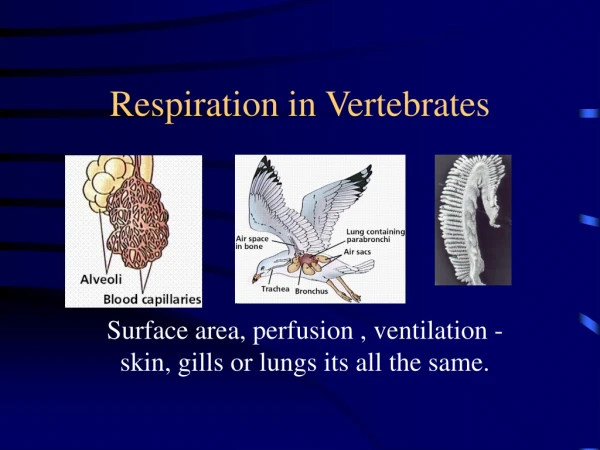
MUSCLES INVOLVED IN RESPIRATION Dr. Ahmed Fathalla Ibrahim & Dr. Zeenat Zaidi
OBJECTIVES At the end of the lecture, students should be able to: • Describe the components of the thoracic cage and their articulations. • Describe in brief the respiratory movements. • List the muscles involved in inspiration and in expiration. • Describe the attachments of each muscle to the thoracic cage and its nerve supply. • Describe the origin, insertion, nerve supply of diaphragm.
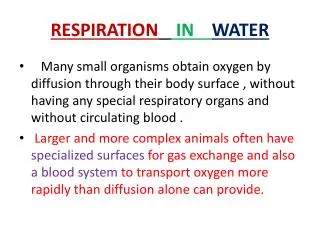
THORACIC CAGE • Conicalin shape • Formed of: • Sternum & costal cartilages: anteriorly • Twelve pairs of ribs: laterally • Twelve thoracic vertebrae: posteriorly • Has 2 apertures (openings): • Superior (thoracic outlet): narrow, open, continuous with neck • Inferior: wide, closed by diaphragm
Articulations Manubriosternal (fibrocartilagenous J.) Small angular Movement possible Costovertebral (Synovial joint) Costochondral (cartilagenous J.) no movements possible Sternocostal (synovial), mobile EXCEPT first, which is cartilagenous & fixed Xiphisternal (fibrocartilagenous J.) no significant movements
Respiratory MovementsA- Movements of Diaphragm Inspiration Contraction (descent) of diaphragm Increase of vertical diameter of thoracic cavity Expiration Relaxation (ascent) of diaphragm)
Respiratory MovementsB- Movements of Ribs PUMP HANDLE MOVEMENT Elevation of ribs Increase in antero-posterior diameter of thoracic cavity BUCKET HANDLE MOVEMENT Elevation of ribs Increase in lateral diameter of thoracic cavity
Inspiratory Muscles • Diaphragm (most important muscle) • External intercostal muscles Rib elevators: • Accessory muscles (only used during forced inspiration): • Scalene muscles: Muscles attaching cervical vertebrae to first & second rib: • Pectoralis major: Muscle attaching thoracic cage to upper limb:
DIAPHRAGM • A musculotendinous partition between thoracic & abdominal cavity • Convex toward thoracic & concave toward abdominal cavity • Origin: • Sternal: xiphoid process of sternum • Costal: lower 6 costal cartilages & 12th rib • From medial & lateral arcuateligaments • Vertebral: as right crusfrom upper 3 lumbar vertebrae & left crus from upper 2 lumbar vertebrae 1 2 Left crus Right crus 3 4 Lateral arcuatelig. Medial arcuatelig.
Insertion: Fibers converge to join the central tendon Nerve supply: phrenic nerve (C3,4,5), penetrates diaphragm & innervates it from abdominal surface Action:contraction (descent) of diaphragm increases vertical diameter of thoracic cavity (essential for normal breathing)
EXTERNAL INTERCOSTAL • Attachments:from lower border of rib above to upper border of rib below • Direction of fibers: downward & medially • Nerve supply: intercostal nerves • Action: rib elevators (inspiratory)
SCALENE MUSCLES Cervical vertebrae • Three muscles: • Scalenus anterior • Scalenusmedius • Scalenus posterior • Origin: cervical vertebrae • Insertion: 1st & 2nd ribs • Action: elevate 1st & 2nd ribs (inspiratory) 2 3 1 1st rib 2nd rib
PECTORALIS MAJOR Origin: sternum + costal cartilages Insertion:humerus Action: increases antero-posterior diameter of thoracic cavity, when arm is fixed (inspiratory)
Expiratory Muscles • Act only during forced expiration • Rib depressors: • Internal intercostal • Innermost intercostal • Subcostals • Transversusthoracis • Anterior abdominal wall muscles: • External oblique • Internal oblique • Transversusabdominis • Rectusabdominis
RIB DEPRESSORS 1. Internal intercostal 2. Innermost intercostal Direction: upward & medially 3. Subcostal 4. Transversusthoracis Nerve supply: intercostal nerves (ventral rami of T1-T11) 3 1 2 4
Anterior abdominal wall • Is formed of 3 layers of muscles of fibers running in different directions (to increase strength of anterior abdominal wall) • The 3 muscles form a tendinous sheath in which a fourth muscles lies (rectus abdominis) • Muscles are attached to: sternum, costal cartilages and ribs + hip bones • The aponeurosis of the 3 muscles on both sides fuse in the midline to form linea alba • Action (during forced expiration): Compression of abdominal viscera to help in ascent of diaphragm • Nerve supply: lower intercostal nerves (T7 – T11), subcostal nerve (T12) and first lumbar nerve. Linea alba EO TA RA IO
Anterior abdominal wall Muscles • Internal oblique (middle layer) Direction: upward & medially External oblique (outer layer) Direction: downward & medially Linea alba
Transversusabdominis(inner layer) Direction: transverse Rectusabdominis Direction:vertical