Download
1 / 28
360 likes | 2.15k Vues
Kingdom of Bahrain Arabian Gulf University College of Medicine and Medical Sciences. Muscles of Respiration. Slides originally prepared by: Dr. Kumar Notes added by: Ali Alhashli. Functions. What are the functions of the thoracic cage? Protects organs and structures of the thorax.

E N D
Kingdom of Bahrain Arabian Gulf University College of Medicine and Medical Sciences Muscles of Respiration Slides originally prepared by: Dr. Kumar Notes added by: Ali Alhashli
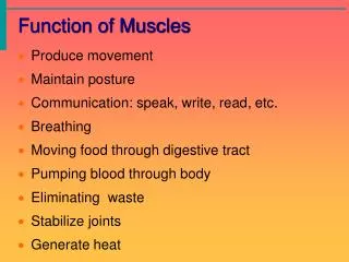
Functions • What are the functions of the thoracic cage? • Protects organs and structures of the thorax. • Provides stable support of upper extremity and head. • Flexible and can change dimensions during ventilation. • What are the functions of thoracic vertebrae? • Protects organs and structure of the thorax. • Protects and houses spinal cord. • Support structures of the thorax and articulate with ribs. • Flexible.
BONES OF THE THORACIC WALL: STERNUM Articulation with clavicle Manubrium Sternal angle (manubriosteral joint) → primary Rib 2 Body of sternum Xiphosternal joint → secondary Xiphoid process Intercostal space Intervertebral discs (Secondary joints) vertebra Costal cartilage
INTERCOSTAL SPACES 1st intercostal space: the space between the first and the second ribs 1 2 • We have 12 pairs of ribs • First seven ribs are true ribs. • Ribs 8-10 are false ribs (they don’t attach directly to the sternum. • Ribs 11 & 12 are floating ribs. 3 4 Transverse ridges 5 6 Interchondral joints (synovial) 11 7 8 12 9 10
INTERCOSTAL SPACE: CONTENTS Skin Intercostal “VAN” in costal groove: Vein, Artery & nerve Superfacial fascia External intercostal muscles Then endothoracic fascia separating the thoracic wall from the parietal pleura of the lung Internal intercostal muscle Deep internal intercostal muscle
INTERCOSTAL MUSCLES EXTERNAL INTERCOSTAL MUSCLES External intercostal Muscles: they run anteroinferiorly and they aid in the process of inspiration External intercostal membrane
INTERCOSTAL MUSCLES EXTERNAL INTERCOSTAL MUSCLES External intercostal membrane External intercostal muscles They run downward and obliquely
INTERCOSTAL MUSCLES INTERNAL INTERCOSTAL MUSCLE S Internal intercostal muscles: they run opposite to External intercostal muscles (postero-inferiorly)
INTERCOSTAL MUSCLES INNERMOST INTERCOSTAL MUSCLES Innermost intercostal muscles: in the same direction of internal intercostal muscles (posterio-inferiorly) and they act with them to aid in the process of expiration
INTERCOSTAL MUSCLES SUBCOSTALES & TRANSVERSUS THORACIS Originating from the inferior body of the sternum and xiphoid process to insert in the costal cartilages of ribs 3-6 Subcostales Transversus Thoracis
RESPIRATORY DIAPHRAGM • Main muscle of inspiration (increasing the vertical volume). • Dome-shaped structure. • Peripheral muscular portion. • Centrally placed tendon of insertion. • Right and Left domes reach as high as the 5th rib (presence of liver and heart). • Right dome slightly higher due to large size of the right lobe of liver.
Parts: 1- Sternal part: from xiphoidprocess 2-Costal part: from lower 6 ribs and their costal cartilages. 3-Vertebral part: from the bodies and discs of lumbar vertebra by means of vertical columns or crura and from the arcuate ligaments.
MUSCLES OF THE THORACIC WALL RESPIRATORY DIAPHRAGM CENTRAL TENDON RIGHT DOME Slightly higher LEFT DOME
RESPIRATORY DIAPHRAGM: Thoracic Surface AORTIC T.V. 12 Right dome VENACAVAL T.V. 8 Left dome OESOPHAGEAL T.V. 10 VOICE OF ARABIC 8, 10, 12 Central tendon
RESPIRATORY DIAPHRAGM (FUNCTIONS) • Functions of the diaphragm: • Muscle of inspiration: Contraction pulls down the central tendon increased vertical diameter of the thorax decreased intrathoracic pressure expansion of the lungs. • Muscle of abdominal straining: by taking a deep breath, closing the glottis, and fixing the diaphragm assists the muscles of the anterior abdominal wall in raising intra-abdominal pressure to evacuate pelvic contents urination, defecation and parturition • Weightlifting muscle: assists in supporting the vertebral column and postvertebral muscles in the lifting of heavy weights. • Thoracoabdominal pump: decreased intrathoracic pressure and increased intraabdominal pressure compression of blood in the inferior vena cava forcing it upward into the heart and also the lymph in the abdominal lymph vessels forcing it upward into the thoracic duct.
Nerve supply of the diaphragm: • Motor: phrenic nerve (which is originating from C3, C4 and C5). • Sensory • Phrenic nerve central portion including parietal pleura and parietal peritoneal covering it. • Lower 6 intercostal nerves peripheral portion
RESPIRATORY DIAPHRAGM: NERVE SUPPLY PHRENIC NERVE (from C3, C4, C5)
PARALYSIS OF THE DIAPHRAGM UNILATERAL PARALYSIS may be due to trauma or surgical division involved half (dome) of diaphragm ascends on inspiration
ACCESSORY MUSCLES OF RESPIRATION Sternocleidomastoid muscle Posterior scalene Middle scalene Anterior scalene
ACCESSORY MUSCLES OF RESPIRATION Pectoralis major Serratus anterior
ACCESSORY MUSCLES OF RESPIRATION ANTERIOR ABDOMINAL WALL MUSCLES Rectus abdominis Internal oblique
Respiratory Movements During inspiration, all 3 diameters increase: 1. Increase in antero-posterior diameter: pump-handle movement of ribs. 2. Increase in transverse diameter: lateral spreading of ribs + bucket-handle movement of ribs. 3. Increase in cranio-caudal diameter: diaphragm
THORACIC CAGE: BOUNDARIES SUPERIOR and INFERIOR Suprapleural membrane (Sibson’s fascia) Diaphragm