Types of Muscles
280 likes | 1.02k Vues
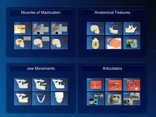
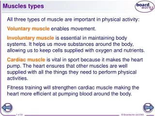
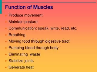
Types of Muscles. Smooth w Involuntary muscle; controlled unconsciously w In the walls of blood vessels and internal organs Cardiac w Controls itself with help from nervous and endocrine systems w Only in the heart Skeletal w Voluntary muscle; controlled consciously

Types of Muscles
E N D
Presentation Transcript
Types of Muscles Smooth w Involuntary muscle; controlled unconsciously w In the walls of blood vessels and internal organs Cardiac w Controls itself with help from nervous and endocrine systems w Only in the heart Skeletal w Voluntary muscle; controlled consciously w Over 600 throughout the body
KEY POINTS w An individual muscle cell is called a muscle fiber. w A muscle fiber is enclosed by a plasma membrane called the sarcolemma. w The cytoplasm of a muscle fiber is called a sarcoplasm. w Within the sarcoplasm, the T tubules allow transport of substances throughout the muscle fiber and the sarcoplasmic reticulum stores calcium.
The Myofibril w Myofibrils are made up of sarcomeres, the smallest functional units of a muscle. w A sarcomere is composed of filaments of two proteins, myosin and actin, which are responsible for muscle contraction. w Myosin is a thick filament with a globular head at one end. w An actin filament—composed of actin, tropomyosin, and troponin—is attached to a Z disk.
Events Leading to Muscle Fiber Action • A motor neuron releases acetylcholine (ACh). 2. ACh binds to receptors on the sarcolemma. 3. The action potential triggers release of Ca2+. 4. The Ca2+ binds to troponin on the actin filament, and the troponin pulls tropomyosin off the active sites, allowing myosin heads to attach to the actin filament.
The Sliding Filament Theory w When myosin cross-bridges are activated, they bind strongly with actin, resulting in a change in the cross-bridge. w The change in the cross-bridge causes the myosin head to tilt toward the arm of the cross-bridge and drag the actin and myosin filaments in opposite directions. w The tilt of the myosin head is known as a power stroke. w The pulling of the actin filament past the myosin results in muscle shortening and generation of muscle force.
Changes in Membrane Potential Depolarization—inside of cell becomes less negative relative to outside (> –70 mV) Hyperpolarization—inside of cell becomes more negative relative to outside (< –70 mV) Graded potentials—localized changes in membrane potential (either depolarization or hyperpolarization) Actionpotentials—rapid, substantial depolarization of the membrane (–70 mV to +30 mV to –70 mV all in 1 ms)
THE NEUROMUSCULAR JUNCTION JUNCTION
Fiber Types • Type I • Type IIa • Type IIb • Type of Muscular Contractions – • Concentric, Eccentric • Isometric, Isotonic, Isokinetic • Origin – Proximal Insertion – Distal
Muscle and Joint Nerve Endings Proprioceptors • Joint kinesthetic receptors in joint capsules sense the position and movement of joints. • Muscle spindles sense how much a muscle is stretched and angle of stretch w Golgi tendon organs detect the tension of a muscle on its tendon, providing information about the strength of muscle contraction.
Chapter 2 • Factors affecting Strength • Amount of Neuromuscular Stimulation • Hypertrophy of Sarcomere • Age (Sarcopenia) • Sex • Angle of Pennation • Prestretching