Download
1 / 34
340 likes | 523 Vues
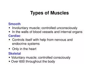
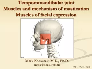
MUSCULI MASTICATORII. Muscles of mastication. 4 pairs of muscles attached to the mandible Movement of temporomandibular joint Arise from the bones of the neurocranium Pennate structure Fasciae Blood supply: maxillary artery Nerve supply: mandibular nerve. Masseter muscle.

E N D
MUSCULI MASTICATORII Muscles of mastication
4 pairs of muscles attached to the mandible • Movement of temporomandibular joint • Arise from the bones of the neurocranium • Pennate structure • Fasciae • Blood supply: maxillary artery • Nerve supply: mandibular nerve
Thick, quadrilateral muscle Superficial and deep portion: The Superficial Portion Origo:maxillary process of zygomatic bone and theanterior ⅔ of the lower border of the zygomatic arch Fibers pass downward and backward Insertion:tuberositas masseterica (the angle and lower ½ of the lateral surface of the ramus of the mandible)
The Deep Portion Smaller and more muscular in texture Origo: posterior ⅓ of lower border and the whole of the medial surface of the zygomatic arch Fibers pass downward and forward Insertion: the upper ½ of the lateral surface of theramus mandible
Functional organization of the human masseter muscle www.springerlink.com/index/U007G453650W2163.pdf
Function • Bilateral contraction: The superficial part: • elevation • propulsion The deep portion: elevation Unilateral contraction: lateropulsion
The Architecture • The typical pennate structure - zones of muscular and aponeurotic attachments • The pennate structure allows spread the infection (submasseteric abscess) • The differential activity of the muscular planes during masticatory function makes it necessary to respect the anatomic and functional individuality in the diagnosis and treatment of dysfunctional disorders of the masticatory apparatus
The Masseteric Fascia • Firmly connected with the muscle • From arcus zygomaticus to basis mandibulae • Dorsally merge with fascia parotis (fascia parotideomasseterica)
Palpation The Superficial Portion The Deep Portion
Broad, triangular muscle Localized in the temporal fossa Origo: - the whole of the temporal fossa - the deep surface of the temporal fascia Fibers converge as they descend, and end in a tendon, which passes deep to the zygomatic arch Insertion: apex and medial surface of the coronoid process (crista temporalis) and the anterior border of the ramus of the mandible to retromolar triangle
Detailed description The temporalis muscle consists of three parts: superficial zygomatic deep portion The human temporalis muscle: Superficial, deep, and zygomatic ... onlinelibrary.wiley.com › ... › Journal Home › Vol 22 Issue 6
The Superficial Portion Origo: temporal aponeurosis, temporal line Insertion: the coronoid process of the mandible
The Zygomatic Portion Origo: superior and medial zygoma and zygomatic arch Insertion: coronoid process Fuse with tendon of the superficial part of the temporalis muscle and with the deep masseter portion. Rostrally interdigitated with the portion of the deep temporalis part
The Deep Portion Origo: bony surface of the frontal, sphenoid, parietal and temporal bones Insertion: coronoid process and the ramus of the mandible, just caudal to the last molar (retromolar triangle) Fibers from deep part interdigitated with the buccinator and the superior pharyngeal constrictor
Function Elevation (anterior part) Retropulsion (posterior part)
The Temporal Fascia Covers the temporal muscle Origo: superior temporal line Insertion: zygomatic arch Has two layers: 1. superficial inserted into the lateral border of the arch 2. deep inserted into the medial border of the arch Between these layers is a small quantity of fat and attachment of the superficial fibres of the temporal muscle
Palpation The Anterior Part The Posterior Part
Short, thick muscle, conical in form Located in the infratemporal fossa Two heads: upper and lower The Upper/Sphenoid Head: Origo: infratemporal facies of the great wing of the sphenoid bone The Lower/Pterygoid Head: Origo: lateral surface of the lateral pterygoid plate
Fibres pass horizontally backward and lateralward Insertion: - fovea pterygoidea (depression in front of the neck of the condyle) - joint capsule and articular disc of the TMJ (front margin)
Alternating tendinous sheets characteristic of a penniform structure
Function Bilateral contraction: propulsion Unilateral contraction: lateropulsion
Thick, quadrilateral Located in the infratemporal fossa Two heads: Origo: - tuberosity of the maxilla (lateralis) - medial surface of the lateral pterygoid plate (medialis) Fibers pass downward, lateralward and backward Insertion: tuberositas pterygoidea (lower and back part of the medial surface of the ramus and angle of the mandible)
1 - lateral part 2 - medial part Inner surface of the ramus mandibulae
The Architecture 7 musculo-aponeurotic layers are making up a penniform structure 7 5 3 1 6 4 2
Function Bilateral contraction: propulsion elevation Unilateral contraction: lateropulsion
The Interpterygoid Fascia Between the medial and lateral pterygoid muscles Origo: the base of the skull Insertion: inner surface of the mandible The border is reinforced by sphenomandibular ligament Contain nerves and vessels Allow spread of infection
pteryg. lat. pteryg. med.