Ch. 5: Integumentary System
Ch. 5: Integumentary System. Integumentary System Functions. Protection chemical: acidic skin secretions, melanin, DNA physical: keratinized cells biological: dendritic cells in epidermis, macrophages in dermis Temperature regulation perspiration, skin blood flow Cutaneous sensation


Ch. 5: Integumentary System
E N D
Presentation Transcript
Integumentary System Functions • Protection • chemical: acidic skin secretions, melanin, DNA • physical: keratinized cells • biological: dendritic cells in epidermis, macrophages in dermis • Temperature regulation • perspiration, skin blood flow • Cutaneous sensation • hairs, and see chapter 13 • Metabolism • synthesizes vitamin D precursor (in presence of sunlight) • Blood reservoir • up to 5% of total blood volume
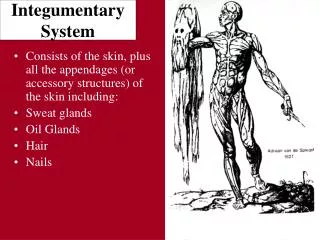
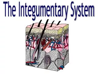
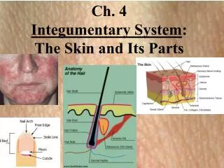
Skin (Integument) • Consists of three major regions • Epidermis – outermost superficial region • Dermis – middle region • Hypodermis (superficial fascia) – deepest region
Epidermis • Composed of keratinized stratified squamous epithelium, consisting of four distinct cell types and four or five layers • Cell types include keratinocytes, melanocytes, Merkel cells, and Langerhans’ cells • Outer portion of the skin is exposed to the external environment and functions in protection
Cells of the Epidermis • Keratinocytes – produce the fibrous protein keratin • Melanocytes – produce the brown pigment melanin • Langerhans’ cells (= epidermal dendritic cells) – epidermal macrophages that help activate the immune system • Merkel cells – function as touch receptors in association with sensory nerve endings
Layers of the Epidermis:Stratum Basale (Basal Layer) • Deepest epidermal layer firmly attached to the dermis • Consists of a single row of the youngest keratinocytes • Cells undergo rapid division, hence its alternate name, stratum germinativum
Layers of the Epidermis: Stratum Spinosum (Prickly Layer) • Cells contain a weblike system of intermediate filaments attached to desmosomes • Melanin granules and Langerhans’ cells (also known as epidermal dendritic cells) are abundant in this layer
Layers of the Epidermis: Stratum Granulosum (Granular Layer) • Thin; three to five cell layers in which drastic changes in keratinocyte appearance occurs • Keratohyaline and lamellated granules accumulate in the cells of this layer
Layers of the Epidermis:Stratum Lucidum(Clear Layer) • Thin, transparent band superficial to the stratum granulosum • Consists of a few rows of flat, dead keratinocytes • Present only in thick skin
Layers of the Epidermis:Stratum Corneum (Horny Layer) • Outermost layer of keratinized cells • Accounts for three quarters of the epidermal thickness • Functions include: • Waterproofing • Protection from abrasion and penetration • Rendering the body relatively insensitive to biological, chemical, and physical assaults
Dermis • Second major skin region containing strong, flexible connective tissue • Cell types include fibroblasts, macrophages, and occasionally mast cells and white blood cells • Composed of two layers – papillary and reticular
Layers of the Dermis: Papillary Layer • Papillary layer • Areolar connective tissue with collagen and elastic fibers • Its superior surface contains peglike projections called dermal papillae • Dermal papillae contain capillary loops, Meissner’s corpuscles, and free nerve endings
Layers of the Dermis:Reticular Layer • Reticular layer • Accounts for approximately 80% of the thickness of the skin • Collagen fibers in this layer add strength and resiliency to the skin • Elastin fibers provide stretch-recoil properties
Hypodermis • Subcutaneous layer deep to the skin • Composed of adipose and areolar connective tissue
Skin Color • Three pigments contribute to skin color • Melanin – yellow to reddish-brown to black pigment, responsible for dark skin colors • Freckles and pigmented moles – result from local accumulations of melanin • Carotene – yellow to orange pigment, most obvious in the palms and soles of the feet • Hemoglobin – reddish pigment responsible for the pinkish hue of the skin
Sweat Glands • Different types prevent overheating of the body; secrete cerumen and milk • Eccrine sweat glands – found in palms, soles of the feet, and forehead • Apocrine sweat glands – found in axillary and anogenital areas • Ceruminous glands – modified apocrine glands in external ear canal that secrete cerumen • Mammary glands – specialized sweat glands that secrete milk
Sebaceous Glands • Simple alveolar glands found all over the body • Soften skin when stimulated by hormones • Secrete an oily secretion called sebum
Hair • Filamentous strands of dead keratinized cells produced by hair follicles • Contains hard keratin which is tougher and more durable than soft keratin of the skin • Made up of the shaft projecting from the skin, and the root embedded in the skin • Consists of a core called the medulla, a cortex, and an outermost cuticle • Pigmented by melanocytes at the base of the hair
Hair Function and Distribution Functions • Maintain warmth • Sensory: alert body to presence of insects on skin • Guard scalp against physical trauma, heat loss, sunlight Distribution: over entire skin surface except: • Palms, soles, lips • Nipples and portions of external genitalia
Hair Follicle • Root sheath extending from the epidermal surface into the dermis • Deep end is expanded forming a hair bulb • A knot of sensory nerve endings (a root hair plexus) wraps around each hair bulb • Bending a hair stimulates these endings, hence our hairs act as sensitive touch receptors
Hair Types • Vellus – pale, fine body hair in children, adult females • Terminal – coarse, long hair of eyebrows, scalp, axillary, and pubic regions Hair thinning, baldness • Alopecia – hair thinning in both sexes • True, or frank, baldness • Genetically determined and sex-influenced condition • Male pattern baldness – due to effect of DHT on follicles
Structure of a Nail • Scalelike modification of the epidermis on the distal, dorsal surface of fingers and toes Figure 5.7
Burns • First-degree – only epidermis is damaged • Localized redness, swelling, and pain • Second-degree – epidermis and upper regions of dermis are damaged • Like first degree burns, but blisters also appear • Third-degree – entire thickness of skin is damaged • Burned area appears gray-white, cherry red, or black; there is no initial edema or pain (since nerve endings are destroyed) • Fourth-degree – entire thickness of skin is damaged • Underlying tissue such as muscle, tendon, ligament also damaged
Rule of Nines • Estimates the severity of burns • Burns considered critical if: • Over 25% of the body has second-degree burns • Over 10% of the body has third-degree burns • There are third-degree burns on face, hands, or feet