Download
1 / 29
360 likes | 692 Vues
The Role of the Major Histocompatibility Complex in the Immune Response. MHC & Immune Response. Molecules coded for by MHC designed to bind peptide fragments from protein antigens, allowing them to be recognized by antigen-specific T cells.

E N D
The Role of the Major Histocompatibility Complex in the Immune Response
MHC & Immune Response • Molecules coded for by MHC designed to bind peptide fragments from protein antigens, allowing them to be recognized by antigen-specific T cells. • MHC molecules = selective, the 3rd set of recognition molecules (TCR & BCR). • Originally recognized by influence on transplantation rejection
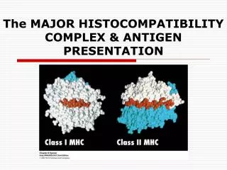
MHC genes encoding three classes of molecules • Class I MHC genes • encode glycoproteins expressed on the surface of nearly all nucleated cells • major function : presentation of peptide antigens to TC cells • Class II MHC genes • encode glycoproteins expressed primarily on antigen-presenting cells (macrophages, dendritic cells & B cells) • major function : presentation of peptide antigens to TH cells • Class III MHC genes • encode various secreted proteins that have immune functions in complement system and inflammation
Similarities between MHC I & II • Function : Both are critical cell interaction molecules & strong transplantation antigens. • Structure : Both are two chain transmembrane proteins, have a single peptide-binding site in the extracellular region unique to a particular allele, have polymorphic & non-polymorphic regions. • Expression : Both are codominantly expressed. • Diversity : Both show genetic polymorphism with multiple alleles in population
Differences between MHC I & II • Cells expressing MHC I + peptide interact with CD8+ T cells; cells expressing MHC II + peptide interact with CD4+ T cells. • MHC I bind peptides (8 - 9 AA) originating from endogenous antigens; MHC II bind peptides (12 - 25 AA) originating from exogenous antigens. • MHC I expression constitutive on most nucleated cells; constitutive MHC II expression is more limited. • MHC II heterodimer coded for entirely with MHC; MHC I contains b2-microglobulin coded for outside MHC
Figure 8.1 Simplified depiction of the human (A) and mouse (B) MHC, showing regions and genes coding for polymorphic MHC class I and II molecules. b2m = b2- microglobulin, encoded outside the MHC.
Figure 8.2 Different depictions of an MHC class I molecule. (A) Diagram of the structures of an MHC class I molecule associated at the cell surface with b2m. (B) Side view of the MHC class I molecule with b2m, showing the peptide-binding groove. (C) Top view of the peptide-binding groove. (D) Diagram of the interaction of a T-cell receptor with an MHC class I molecule and peptide bound in the peptide-binding groove. [Figures B and C from Bjorkman et al., 1987, with permission; Figure D adapted from Rammensee et al., 1993.]
Figure 8.3 Different depictions of an MHC class II molecule. (A) Diagram of the struc- [cl11]ture of an MHC class II molecule at the cell surface. (B) Side view of the MHC class II molecule showing the peptide-binding groove. (C) Top view of the peptide-binding groove. [Adapted from Stern et al., 1994, with permission.] (D) Diagram of the interaction of a T-cell receptor with an MHC class II molecule and peptide bound in the peptide-binding groove.
Antigen processing & presentation (“rules”) • Protein must be degraded to fragments to bind to MHC. • Association between MHC & peptides are selective. • MHC I bind to cytosolic peptides, whereas II from outside of cells • Peptide breakdown occurs in two places: 1) within cytoplasm & ER --- bind to MHC I, 2) within acid vesicles --- bind to MHC II • Binding affinity is similar to Ab/Ab interaction • Peptide-MHC complex forms, and dissociate slowly. • Self-MHC restriction of T cells (next figures) • Both CD4+ & CD8+ T cells can recognize antigen only when it is presented with self-MHC on the membrane of another.
Experimental demonstration that antigen processing is necessary for TH-cell activation
Figure 8.4 Processing of an exogenous antigen in the MHC class II pathway. Ii = invariant chain, CLIP = fragment of Ii bound to MHC class II groove.
Figure 8.5 Selective binding of processed peptides by different MHC alleles. The numbers refer to positions of amino acids in the sequence of the protein antigen.
Figure 8.6 Processing of an endogenous antigen in the MHC class I pathway. b2m = b2-microglobulin.