Download
1 / 36
380 likes | 421 Vues
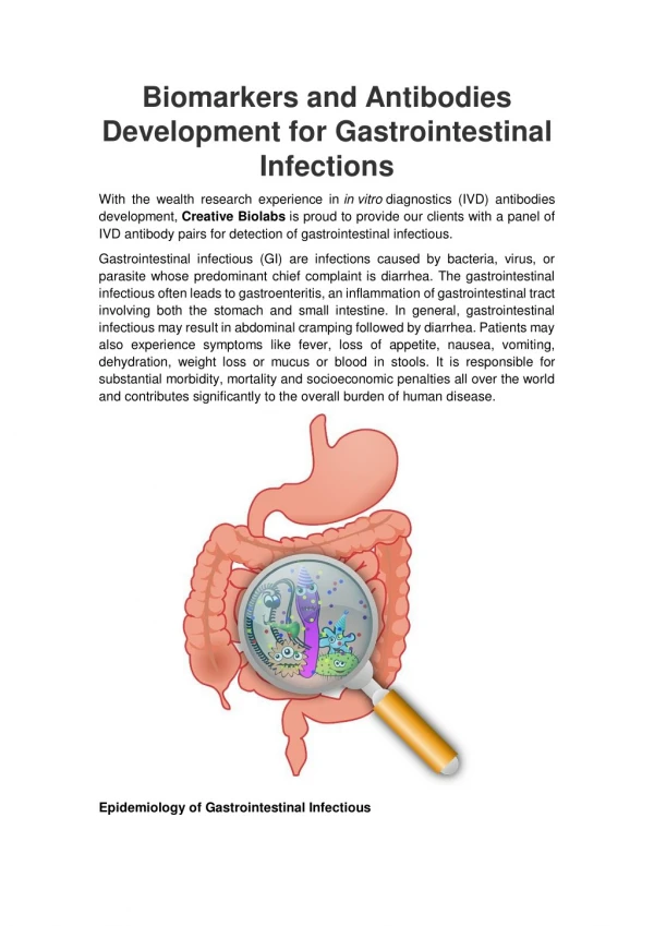
Explore the fascinating world of gastrointestinal infections, from bacterial overgrowth to pathogenic microorganisms causing diarrhea and dysentery. Learn about bacterial toxins, enteroinvasive E. coli strains, and the distinctive characteristics of Salmonella infections.

E N D
Topic 04Gastrointestinal infections II Lecture for 3rd-year students General medicine Ondřej Zahradníček (with use of prof. Votava‘s last year sources) 23rd of October, 2015
Revision 1 • Gastrointestinal tract is an area interesting from microbiology side of view • It contains two important ecosystems, where bacteria are normally found and use of antibiotic is often contraproductive: • Oral cavity with typical biofilm system (supragingival and subgingival plaque) • Intestine (especially large intestine) with about 1 kg of microbes, mostly anaerobic • On the other hand, other areas contain less or no microorganisms (oesophagus, stomach…)
Revision 3 *but starting in the intestine
Revision 4: Intestinal syndromes Bacterial overgrowth syndrome: after surgery, during depressed peristalsis or gastric achlorhydria bacteria may overgrow in the small intestine → steatorrhoea, deficiency of vitamin B12, diarrhoea, malabsorption of vitamins A and D Diarrhoea: increase in daily amount of stool water – common intestinal response to many agents Dysentery: acute inflammation of the colon → abdominal pain & small-volume stools with blood, pus and mucus
Revision 4: Intestinal pathogens Bacteria: they may cause Intestinal infection (e. g. salmonellosis) Enterotoxicosis (staphylococcal, Bacillus cereus etc.) Viruses: quite common, often not diagnosed Fungi: present normally in the intestine, but sometimes overmultiplied Parasites: not always causing diarrhoea, sometimes it can be even obstipation
Enterotoxicosis (food poisoning) The term „food poisoning“ is commonly used for different situations, including infections. Nevertheless, it is necessary to understand that diarrhoea related with bacteria can be 1) Enterotoxicosis = intoxication due to a toxin produced by bacteria in the food; there is no incubation • Staphylococcus aureus enterotoxicosis (a superantigen) • Clostridium perfringens: heat-labile enterotoxin • Bacillus cereus: heat-stable enterotoxin and vomiting toxin (mostly in rice) • Clostridium botulinum: heat-labile neurotoxin 2) Infection caused by bacteria that produce toxins inside the intestine (e. g. enterotoxic strains of Escherichia coli); these are not considered an enterotoxicosis, incubation period exists 3) Infection caused by bacteria that do not produce toxins, but are invasive (e. g. enteroinvasive strains of Escherichia coli)
Enteric infection caused by E. coli Escherichia coli strains causing diarrhoeal disease: • ETEC (enterotoxic E. coli): children in developing countries, traveller´s diarrhea; 2 enterotoxins (heat-labile and heat-stable) • EPEC (enteropathogenic E. coli): approx. 12 serotypes, for example O55, O111; small infants (< 3yo); disruption of microvillus structure; E. coli strains are tested for EPEC • EIEC (enteroinvasive E. coli): similar to Shigella invasion → dysentery-like disease; invasion of colonic cells • STEC (shigatoxigenous E. coli): also known as VTEC (verotoxigenous E. coli); for more information see further • EAggEC (enteroaggregative E. coli) and other pathogenicity groups Do not forget that majority of E. coli strains are „normal“ strains helping us by production of vitamins etc.!
STEC/VTEC – more information • those strains produce 2 cytotoxic shigatoxins • mechanism of pathogenesis: destruction of microvilli • hemorrhagic colitis & hemolytic-uremic syndrome (HUS) • HUS – three typical signs: • so called mikroangiopatic anemia • trombocytopenia (lack of platelets) • acute renal failure • Letality in HUS is 5 %, almost in children < 5 yo; often lasting ill effects • in 2011 there was a strain O104:H4 in Gerrmany, more common is strain O157:H7 • subgroup of STEC is EHEC (enterohaemorrhagic E. coli)
Escherichia coli – good and bad www2.mf.uni-lj.si/~mil/bakt2/bakt2.htm
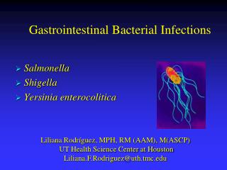
Salmonella infections Two types of salmonella infections: 1) Systemic infections caused by anthropopathogenic serovarsS. Typhi, S. Paratyphi A–C; the intestine is just gate of infection 2) Intestinal infections caused by zoopathogenic serovarsS. Enteritidis, S. Typhimurium, S. Infantis etc.; there exist more than 4000 serotypes; in some cases even these may cause systemic infections, but in majority of them it is just intestinal infection Pathogenesis of both starts with the invasion of intestinal epithelia In 1) invasion continues and infection becomes generalized → little or no diarrhoea, but pronounced fever & other general symptoms; common carriership even for many years! In 2) infection is localized to ileocaecal region → diarrhoea, nausea & vomiting, abdominal pain, temperature may be elevated
Salmonela on MAL agar Foto O. Z.
Typhoid fever – diagnostics and treatment Diagnostics and treatment of diarrhoeic infections will be described later, so here only for Typhoid fever: Diagnostics: Direct diagnostics – specimens of blood, urine (!) and stool tested for Salmonella, serotyping for S. Typhi Indirect diagnostics – in later phases – Widal reaction (agglutination in test tubes) Treatment: antibiotics (chloramphenicol, fluorochinolones, ampicillin, cotrimoxazol)
Campylobacter and Shigella Campylobacter jejuniintestinal infections are recently even more frequent than salmonella infections; the microbe invades jejunal epithelium. Poultry is the reservoir Shigella is a bacterium that does not exist according to recent genetic findings (it is just a „special case of E. coli). Nevertheless it is still considered existing for practical reasons. It contains four species: Shigella sonnei, S. flexneri, S. boydii, S. dysenteriae • Very low infectious dose → epidemic outbreaks • Transmitted only among humans • Invasion of cells of colon and rectum; in some strains also shiga toxin (the same as in STEC strains of E. coli) • The disease is called bacterial dysentery
Campylobacter jejuni www.cdc.gov/ncidod/eid/vol5no1/altekruseG.htm.
Yersinia and Vibrio Yersinia enterocolitica causes gastroenteritis, in children also mesenterial lymphadenitis (mimicking acute appendicitis). Contaminated food is a vehiculum of infection; the microbe multiplies in refrigerator even at 4 °C Vibrio cholerae acts through cholera toxin. This toxin activates adenylate cyclase → hypersecretion of water & electrolytes → death by dehydration and electrolyte abnormalities. V. cholerae flourishes in water & causes epidemics. Most important serovars are O1 (with biotypes Classis and El Tor) and O139 Vibrio parahaemolyticus:from raw fish & shell-fish
Vibrio cholerae http://www.cs.dartmouth.edu/brd/Research/Bio/water-borne-bioterrorism.htm
Post-antibiotic diarrhoea Use of antibiotics makes considerable changes in the intestinal ecosystem; many parts of normal flora are destroyed and some other multiply much more than normally Clostridium difficile (CLDI) infection: after any antibiotic, but most frequent after lincosamides (lincomycin or clindamycin) • dangerous pseudomembranous colitis • patients contaminate the hospital environment with resistant spores • diagnostics and treatment: see next slide Infections due to other bacteria: common after tetracyclines; from excessively multiplied Staphylococcus aureus, Pseudomonas aeruginosa or also Candida albicans (an example of diarrhoea of mycotic origin)
CLDI infections: diagnostics • Diagnostics: stool is sent to the laboratory (stool, not rectal swab) and examined for CLDI toxin and CLDI antigen using an immunochromatography test. Possible results: • Toxin +, antigen + means clear infection • Toxin –, antigen + means that infection is still possible (the toxin detection might be false negative) • Toxin –, antigen – means that the infection is very unlikely
CLDI infections: treatment • Treatment: • metronidazol (milder cases) • vancomycin taken orally (!) (severe cases) and recently also fidaxomycin (new and very expensive drug) • it is also possible to use so called faecal bacteriotherapy – a stool donor (usually a relative) is tested for many infections; the stool is then mixed with saline and the „cocktail“ is usually administered using a nasogastric sond
Viral agents of diarrhoea Generally: the most typical agents are small, acid- and bile-resistant non-enveloped viruses Rotaviruses (Reoviridae family) cause serious diarrhoea of young children, epidemics in winter Noroviruses and sapoviruses (formerly agents Norwalk and Sapporo, Caliciviridae family) epidemics in children and adults, too; norovirus infections are quite common, especially in maritime countries; seefood is common source and people travelling on oversea ships are often infected Astroviruses (star-shaped virions) Adenoviruses type 40 and 41 Small, round gastroenteritis viruses
Rotavirus http://web.uct.ac.za/depts/mmi/stannard/emimages.html
Parasitic agents of diarrhoea In previously healthy individuals: Entamoeba histolytica:amoebic dysentery • Giardia lamblia: giardiasis • Cryptosporidium parvum: cryptosporidiosis • Cyclospora cayetanensis In AIDS patients also: • Isospora belli (coccidium) • Strongyloides stercoralis hyperinfection (helminth)
Giardia intestinalis CD-ROM „Parasite-Tutor“ – Department of Laboratory Medicine, University of Washington, Seatle, WA
Other intestinal parasites not causing diarrhoea (helminths) Small intestine: • Ascaris lumbricoides (human roundworm) • Ancylostoma duodenale (Old World hookworm) • Necator americanus(New World hookworm) • Strongyloides stercoralis (threadworm) • Fasciolopsis buski (giant intestinal fluke) • Taenia saginata (beef tapeworm) • Taenia solium (pork tapeworm) • Hymenolepis nana (dwarf tapeworm) • Diphyllobothrium latum (fish tapeworm) Large intestine: • Enterobius vermicularis (pinworm) • Trichuris trichiura (whipworm)
Ascaris lumbricoides (roundworm) egg CD-ROM „Parasite-Tutor“ – Department of Laboratory Medicine, University of Washington, Seatle, WA
Fungi and diarrhoea Candida albicans and other members of Candida genus are normally present in the intestine, but overmultiplying (after antibiotic treatment or radiotherapy) causes infections. Treatment is, nevertheless, also indicated in case of extraintestinal (commonly vaginal) infections with intestinal reservoir Enterocytozoon bieneusi(microsporidium) is a causative agent in diarrhoea in AIDS patients. (Microsporidia is a strange group of fungi, formerly considered to be parasites)
Candida albicans http://academics.hamilton.edu/biology/kbart/image/candida.jpg
Intestinal infections × intestinal transmission • Not all faecal-oral route transmitted infections are intestinal infections. Example: poliomyelitis attacks neural system, but is faecal-orally transmitted • On the other hand not all intestinal infections are strictly faecal-orally transmitted • Faecal oral transmission has several subtyptes • alimentary (food contamination; infections with big infectious dose, e. g. salmonellosis) • dirty hands and fomites (shigellosis) • passive vectors (flies, cocroaches)
Diarrhoea treatment • Diarrhoea treatment is not related to the causative agent (with exception of parazital diarrhoea, where antiparazital drugs are used) • Main goal is water supply and care about organism • Antibiotic treatment is not used even in bacterial diarrhoea, the effect to the infection is weak, but they paradoxically prolonge the positivity for an infection (e. g. for Salmonella) • Exceptions: travellers (it is necessary to solve it), very serious infections (E. coli O104:H4); quinolones or some other drugs are used • Other treatment: charcoal, eventually drugs with only topic antimicrobial activity
Microflora care • In convalescence after diarrhoea, but also after systemic antibicrobial therapy we try to renew normal microflora • We use yogurt (non-sweet, non-fat), fermented cabbage, drugs (Hylac) • Some of them contain substrates for „good“ bacteria, these are prebiotics. • Some containg directly the bacteria, those are probiotics • Some contain both, those are symbiotics
Prevention of intestinal infections • Care about water sources • Good food hygiene (farms, stores, but also our kitchens!) • No cross contamination (do not store already prepeared meals with fresh eggs or meat) • Personal hygiene (children education) • Fighting passive vectors (flies, other insects) • Public health regulations in persons positive for some infections (no work with food, no attendance of school etc.)
How to také stool for individual examinations • Bacteria – Amies (or similar) transport medium • Yeasts – Amies transport medium, eventually a special fungal transport medium (FungiQuick) • Viruses – a hazel-nut sized specimen • Parasites – a hazel-nut sized specimen, traveller anamnesis, usually three specimens • CLDI toxin – a hazel-nut sized specimen • Threadworms – Graham method (see practical sessions)
Diagnostics of bacterial agents • Microscopy of specimen is usually not used • Cultivation uses specific media, almost for enterobacteria(see next slide) • Direct examination of toxins Aa B(Clostridium difficile) and its antigen Diagnostics of viral agents: usually antigen detection, eventually DNA detection Diagnostika parasital and fungal agents:special parasitonogy and mycology methoda www.oxoid.cz
Stool processing Day 0. (sampling) Negative result – after 48 h Positive result – 72 h and more 24 h selenit XLD Endo MCs KA 28°C 42°C 48 h MAL Endo NaCl CIN CCDA Selenit,e XLD, MAL – for Salmonella diagnostics CIN – Yersinia CCDA – Campylobacter NaCl – staphylococci MCS – some STECs Endo – Enterobacteria Blood agar – some more + 72hod identifikace *If not mentioned specially, culture is performed at 37°C
Thanks for listening http://www.giantmicrobes.com/images/doll/salmonella.jpg