Download
1 / 26
280 likes | 564 Vues
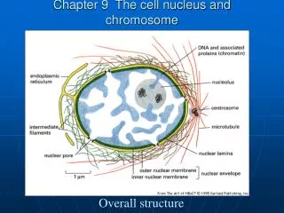
Basics of protein structure and stability IV: Anatomy of protein structure continued. Biochem 565, Fall 2008 09/03/08 Cordes. the main-chain can hydrogen bond to itself. there are also side-chain acceptors and donors. the carbonyl oxygen: main-chain hydrogen bond acceptor.

E N D
Basics of protein structure and stability IV: Anatomy of protein structure continued Biochem 565, Fall 2008 09/03/08 Cordes
the main-chain can hydrogen bond to itself there are also side-chain acceptors and donors the carbonyl oxygen: main-chain hydrogen bond acceptor the amide nitrogen: main-chain hydrogen bond donor
Hydrogen bond geometry • Hydrogen bond not really a covalent ”bond”--not much orbital overlap. • Model as an electrostatic interaction between two dipoles consisting of the H-N bond and the O sp2 lone pair. In electrostatic theory, the optimal orientation of two such dipoles is head-to-tail. The energy of such an arrangement should decrease as the head and tail are brought together as long as atomic van der Waals radii are not violated (then repulsive forces quickly take over). • “Ideal” hydrogen bond in this model would haver~3.0 Å, p=180°, b=0° and g=±60°. Convince yourself of this. • In small molecule crystals, this is approximately what is observed, though there is a lot of variation in the angles b and g. Thus the precise C=O…H angle parameters are not critical. • Main chain-main chain hydrogen bonds found in proteins will show various deviations from this geometry, partly due to the topological constraints imposed by forming secondary structures.
What is a “reasonable” hydrogen bond? Criteria for identifying hydrogen bonds are somewhat arbitrary and many have been used. Here are a couple of examples. • Geometric criteria: Often H-bonds are just identified by two parameters, the O…N (acceptor-donor) distance r, and a O…H-N angle p. The angles describing the C=O…H geometry are sometimes ignored. Typical cutoffs: p > 120° and r < 3.5 Å. (Baker & Hubbard, 1984) • Electrostatic criteria: One of the most commonly used criteria is a potential function based on a pure electrostatic model (Kabsch & Sander, 1983). Place partial positive and negative charges on the C,O (+q1,-q1) and N,H (+q2,-q2) atoms and compute a binding energy as the sum of repulsive and attractive interactions between these four atoms: E=q1q2(1/r(ON)+1/r(CH)-1/r(OH)-1/r(CN))*f where q1=0.42e and q2=0.20e, f is a dimensional factor (=332) to convert E to kcal/mol, and r(AB) is the interatomic distance between atoms A and B. A hydrogen bond is then identified by a binding energy less than some arbitrary cutoff, e.g. E< -0.5 kcal/mol. • Note that the criteria defined above are only applicable when hydrogen atom positions are available. Crystal structures do not have hydrogens--however, their positions can be computed in many cases.
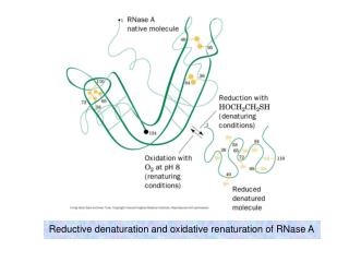
Identifying main-chain H-bonds in X-ray structures of proteins X-ray structures of proteins do not in general include hydrogen atom coordinates--get used looking at pictures of proteins without the hydrogens, and having your mind fill them in Presta LG & Rose GD Science 240, 1632 (1988)
Secondary structure elements in proteins reflect the tendency of backbone to hydrogen bond with itself in a semi-ordered fashion when compacted A secondary structure element is a contiguous region of a protein sequence characterized by a repeating pattern of main-chain hydrogen bonds and backbone phi/psi angles alpha-helix (local interactions) beta-strand(nonlocal interactions)
Local backbone H-bonding: DSSP turn/helix definitions Kabsch & Sander, 1983 3-turn: ‘>’ ‘3’ ‘3’ ‘<‘ notation -N-C-C--N-C-C--N-C-C--N-C-C- residues H O N O H O H O >----------------< H-bond 4-turn: ‘>’ ‘4’ ‘4’ ‘4‘ ‘<‘ notation -N-C-C--N-C-C--N-C-C--N-C-C-N-C-C residues H O N O H O H O H O >----------------------< H-bond 5-turn (just an elaboration of 3- and 4-turn. A minimal helix is two consecutive N-turns-- for a minimal four helix from residue i to i+3: i <--residue >444< and >444< overlap to give >4444< which defines a helix HHHH from i to i+3 ‘H’ is the notation for a residue in a 4-helix. Notice that the helix does not include the residues involved in the terminal H-bonds. Longer helices are overlapping minimal helices.
the alpha-helix: repeating i,i+4 h-bonds 11 10 12 right-handed helical region of phi-psi space 9 8 7 5 6 4 hydrogen bond 1 By DSSP definitions, which of residues 1-12 are in the helix? Does this coincide with the residues in the helical region of phi-psi space? 3 2
The a-helix, with i,i+4 h-bonds, is not the only way to have local hydrogen bonding of the backbone to itself. The 310 helix has hydrogen bonds between residues i and i+3 The p helix has hydrogen bonds between residues i and i+5. For a number of reasons almost all helices in proteins are a-helices--include backbone, side chain steric issues, van der Waals contacts, H-bond geometry 310 helix a-helix p helix these are poly-Ala, so the gray balls on the outside are b-carbons from the side chains
310 and helices have sterically allowed conformations but not in the most favored regions of phi-psi space
Helix nomenclature: a-helix example • Hydrogen bond between C=O H-N (residue i) (residue i+4) 15 helix • Repeating unit: • 5 turns • 18 residues per repeat 185 helix • Loop formed between C=O H-N • 13 atoms 1 5 • 3.6 residues per turn 3.613 helix nomenclature used for 310 helix • helices extend with approximately 1.5 Angstrom per residue, 5.4 Angstrom per turn.
Nonlocal backbone hydrogen bonding:DSSP bridge, ladder and sheet definitions parallel bridge: ‘x’ notation -N-C-C--N-C-C--N-C-C- residues H O H O H O \ . . / H-bonds \. ./ (\ and /, .\ /. or .) . \ / . H O H O H H residues -N-C-C--N-C-C--N-C-C- ‘x’ notations ladder= set of one or more consecutive bridges of identical type sheet= set of one or more ladders connected by shared residues antiparallel bridge: ‘X’ notation -N-C-C--N-C-C--N-C-C- residues H O H O H O . ! ! . H-bonds . ! ! . (! or .) . ! ! . . ! ! . O H O H O H residues -C-C-N--C-C-N--C-C-N- ‘X’ notations Kabsch & Sander, 1983
beta strands/sheets beta-strand region of phi-psi space 57 56 54 53 52 51 Is this a parallel or anti-parallel sheet? 50 By DSSP definitions, which of res 49-57 are in the sheet? Does this coincide with the residues in the beta-strand region of phi-psi space? 49
Principal types of secondary structure found in proteins y Repeating (f,y) values a-helix (15) (right-handed) -63o -42o 310helix (14) -57o -30o Parallel b-sheet -119o +113o Antiparallel b-sheet -139o +135o
antiparallel beta-sheet parallel beta-sheet All the most common secondary structure conformations fit nicely within sterically allowed regions of phi-psi space alpha-helix
Because of the repetitive nature of secondary structures, and particularly beta-sheets, proteins can form fibrillar structures and aggregates amyloid-like fibril(left) of peptide GNNQNNY from the yeast prion protein Sup35, and itsatomic structure (right) fibril axis in the case of this fibril the side chains also hydrogen bond to each other amide stacks Nelson et al (Eisenberg lab), Nature 435:773 (2005). for background on “polar zippers”: Perutz et al. PNAS 91:5355 (1991) These types of fibrils important in Huntington’s disease etc
Fibrillar helical structures: the leucine zipper Leu Leu The GCN4 dimer is formed through hydrophobic interactions between leucines (red) in the two polypeptide chains GCN4 “leucine zipper” (green) bound as a dimer (two copies of the polypeptide) to target DNA
Are main-chain H-bonds why proteins are special? “It would seem extraordinary that no other polymer structures exist in which internal hydrogen bonding can give rise to periodically ordered conformations, but no others have been found thus far. We are therefore forced to recognize the uniqueness of this capacity in polypeptide chains, one which enables them to meet the exacting and sophisticated demands of structure and function” --Doty P, Gratzer WB in Polyamino acids, polypeptides and proteins, pp. 111-118, 1962, University of Wisconsin Press see also Honig F & Cohen FE Folding & Design1, R17-20 (1996).
Globular proteins • Keep in mind, however, that if that were all proteins could do, they would just form regular repeating structures. Instead many proteins have globular structures consisting of short secondary structure elements connected by loops and turns that are not necessarily characterized by repeating hydrogen bond structures, but which serve in part to reverse the direction of the polypeptide chain. loops and turns
b-turns in proteins:reversing the chain direction turn residues A b-turn consists of two residues, where there is a hydrogen bond between the carbonyl of the residue preceding the turn and the amide nitrogen following the turn. There are a number of ways to configure the backbone to achieve this. direction ofpolypeptide chain
four basic tight b-turns that all yield an i,i+3 hydrogen bond [from Wilmot CM & Thornton JM J Mol Biol 203, 221(1988)]
Side chain conformation • side chains differ in their number of degrees of conformational freedom (some don’t have any, such as Ala and Gly) •but side chains of very different size can have the same number of c angles.
Side chain conformations--canonical staggered forms Newman projections for c1 of glutamate: glutamate t=trans, g=gauche name of conformation Side chain angles are defined moving outward from the backbone, starting with the N atom: so the c1 angle is N–Ca–Cb–Cg, the c2 angle is Ca–Cb–Cg –Cd ... IUPAC nomenclature: http://www.chem.qmw.ac.uk/iupac/misc/biop.html
Rotamers • a particular combination of side chain torsional angles c1, c2, etc. for a particular residue is known as a rotamer. • for example, for leucine, if one considers only the canonical staggered forms, there are nine (32) possible rotamers: g+g-, g+g+, g-g-, g-g+, tg+, g+t, tg-, g-t, tt • not all rotamers are equally likely. • for example, valine prefers its t rotamer (picture at right) distribution of valine rotamers in protein structures (from Ponder & Richards, 1987) c1=180°, trans or t c1=0 180 360
Side chain rotamers are not limited to canonical eclipsed forms--there are many subtly different rotamers This figure simply shows that the more structures you examine, the more different rotamers become apparent--so as databases of structure have increased, so has the richness of our understanding of side chain conformation. from Xiang & Honig, 2001 How many rotamers there are also depends on how you define whether two conformations represent different rotamers: An “x degree rotamer” in this figure means that at least one side chain angle differs by x degrees: hence classifying rotamers by a 10 degree difference standard is finer grained than classifying them by, say, a 40 degree standard