Download
1 / 12
120 likes | 154 Vues
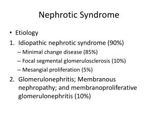
On the basis of observational studies, the most common cause of nephrotic syndrome in school aged children is minimal change disease. On the basis of research evidence and consensus, corticosteroids are considered first line therapy for treatment of nephrotic syndrome. On the basis of consensus, prednisone therapy should be initiated at doses of 60 mg m2 per day 2 mg kg per day administered for 4 to 6 weeks, followed by 40 mg m2 per dose 1.5 mg kg every other day for at least 6 to 8 weeks. On the basis of consensus and expert opinion, it is important to recognize and manage the complications that can arise in patients with nephrotic syndrome, such as dyslipidemia, infection, and thrombosis. On the basis of research evidence, consensus, and expert opinion, several alternative therapies have been observed to have variable efficacy in children with both corticosteroid dependent and corticosteroid resistant nephrotic syndrome, although caution must be exercised in the administration of these corticosteroid sparing medications secondary to toxic adverse effects. On the basis of observational studies, the course of nephrotic syndrome in most patients is that of relapse and remission. Savita Sahu "Basics of Nephrotic Syndrome" Published in International Journal of Trend in Scientific Research and Development (ijtsrd), ISSN: 2456-6470, Volume-3 | Issue-2 , February 2019, URL: https://www.ijtsrd.com/papers/ijtsrd21436.pdf Paper URL: https://www.ijtsrd.com/other-scientific-research-area/other/21436/basics-of-nephrotic-syndrome/savita-sahu<br>

E N D
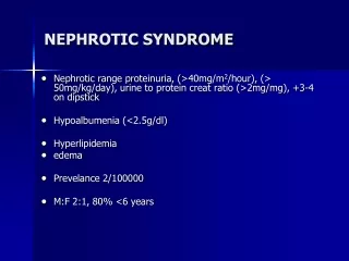
International Journal of Trend in Scientific Research and Development (IJTSRD) Volume: 3 | Issue: 2 | Jan-Feb 2019 Available Online: www.ijtsrd.com e-ISSN: 2456 - 6470 Basics of Nephrotic Syndrome Savita Sahu B.SC Dialysis Technology, Pt. J.N.M. Medical College, Raipur, Chhattisgarh, India ABSTRACT On the basis of observational studies, the most common cause of nephrotic syndrome in school-aged children is minimal change disease. On the basis of research evidence and consensus, corticosteroids are considered first-line therapy for treatment of nephrotic syndrome. On the basis of consensus, prednisone therapy should be initiated at doses of 60 mg/m2 per day (2 mg/kg per day) administered for 4 to 6 weeks, followed by 40 mg/m2 per dose (1.5 mg/kg) every other day for at least 6 to 8 weeks. On the basis of consensus and expert opinion, it is important to recognize and manage the complications that can arise in patients with nephrotic syndrome, such as dyslipidemia, infection, and thrombosis. On the basis of research evidence, consensus, and expert opinion, several alternative therapies have been observed to have variable efficacy in children with both corticosteroid-dependent and corticosteroid-resistant nephrotic syndrome, although caution must be exercised in the administration of these corticosteroid-sparing medications secondary to toxic adverse effects. On the basis of observational studies, the course of nephrotic syndrome in most patients is that of relapse and remission. Keywords: nephrotic syndrome, odema, dyslipidemia INTRODUCTION ?Nephrotic syndrome may occur when the filtering units of the kidney are damaged. This damage allow protein normally kept in the plasma to leak into the urine in large amount. Which reduced the amount of protein in the body. ?Since the protein in the blood help keep fluid in the bloodstream, some of this fluids leaks out of the blood streaminto the tissue, causing swelling, called oedema. ?The swelling may be most noticeable in the legs after have been standing and around the eyeswhen first getup in the morning. Eventually the swelling in the legs may be there all the time and it may also occur in other parts of the body. ?MCNS is one of the causes of primary or idiopathic nephrotic syndrome, to be distinguished from the secondary causes of nephrotic syndrome, such as diabetic nephropathy or amyloidosis. Overall the incidence of primary or idiopathic nephrotic syndrome, including MCNS and its variant. ?When proteinuria is sever it causes the nephrotic syndrome, less sever proteinuria or severproteinuria of short duration, may be asymptomatic. ? The most specific microscopic urinalysis finding is the presence of oval fat bodies. These are sloughed tubular epithelial cells that have reabsorbed some of the excess lipids and lipoproteins in the urine. ?Nephrotic syndrome results from a problem with the kidneys filters, called glomeruli. Glomeruli are tiny blood vessels in the kidneys that remove wastes and excess fluids from the blood and send them to the bladder as urine. As blood passes through healthy kidneys, the glomeruli filter out the waste products and allow the blood to retain cells and proteins the body needs. ?Nephrotic syndrome is a kidney disease with proteinuria, hypoalbuminemia and oedema. Nephrotic- range proteinuria is 3 grams per day or more. On a single spot urine collection, it is 2 g of protein per gram of urine creatinine. ? There are many specific cause of nephrotic syndrome. These include kidney diseases such as minimal changes nephropathy, focal glomerelosclerosis and membranous nephropathy. Nephrotic syndrome can also result from systemic diseases that affect other organs in addition to the kidneys, such as diabetes, amyloidosis and lupus erythematous. ?Nephrotic syndrome (NS) is a condition that is often caused by any of a group of diseases that damage the kidneys' filtering system, the glomeruli. The structure of the glomeruli prevents most protein from getting filtered through into the urine. Normally, a person loses less than 150 mg of protein in the urine in a 24-houre period. ?Nephrotic-range proteinuria, the urination of more than 3.5 grams of protein during a 24-hour period, or 25 times the normal amount, is the primary indicator of NS. Nephrotic syndrome is a kidney disorder that causes your body to excrete too much protein in your urine. ? Nephrotic syndrome is usually caused by damage to the clusters of small blood vessels in your kidneys that filter waste and excess water from your blood. Nephrotic syndrome causes swelling (oedema), particularly in your feet and ankles, and increases the risk of other health problems. ?Treatment for nephrotic syndrome includes treating the underlying condition that's causing it and taking medications. Nephrotic syndrome can increase your risk of infections and blood clots. Your doctor may recommend medications anddietary changes to prevent these and other complications of nephrotic syndrome. DEFINITION 1.Nephrotic syndrome is a nonspecific kidney disorder characterized by a number of signs of disease, proteinuria, hypo-albuminemia and oedema. It is characterized by an increase in permeability of the capillary wall of the Glomerular leading to the presence of high level of protein passing from the blood into the urine. 2.Nephrotic syndrome is a collection of symptoms that indicate kidney damage. Nephrotic syndrome includes following:- @ IJTSRD | Unique Reference Paper ID – IJTSRD21436 | Volume – 3 | Issue – 2 | Jan-Feb 2019 Page: 591
International Journal of Trend in Scientific Research and Development (IJTSRD) @ www.ijtsrd.com eISSN: 2456-6470 ?Proteinuria- large amount of protein in the urine. ?Hyperlipidaemia- higher than normal fat and ?Cholesterol level in the blood. ?Oedema, or swelling usually in the legs and feet, or ankles andless often in the hand or face. ?Hypoalbuminemia- low levels of albumin in the blood. 3.Kidney disease, especially when characterized by oedema and theloss of protein from the plasma into the urine due to increased glomerular permeability is called nephrotic syndrome. 4.Nephrotic syndrome is a characterised by:- ?Proteinuria (24hr urine)> 3.5 g/day. ?40 mg/m2/hr. ?Hypoalbuminemia< 2.5 g/dl. ?Oedema. ?Orbital. ?Pitting. ?Dyslipidaemia. ?Frothy urine. 5.Nephrotic syndrome is an abnormal condition that is marked especially by deficiency of albumin in the blood and excess excretion of protein in the urine due to altered permeability of the glomerular basement membranes.(as from membranous glomerulonephritis, minimal change disease, or diabetes mellitus. 6.Nephrotic syndrome is define as present some clinical feature like: Oedema, urine protein:creatinine ratio ≥2000 mg/g; urine protein ≥300 mg/dL, dipstick urine protein 3+, hypoalbuminemia ≤2.5mg/l. (KDOGI guideline 2012) KDIGO Definitions in Adult FSGS: S. No. Classification Definition <0.3 g/d (<300 mg/g) and normal serum creatinine and serum albumin >3.5 g/dL (35 g/L) 0.3–3.5 g/d (300–3500 mg/g) and change in serum creatinine <25% or 0.3–3.5 g/d (300–3500 mg/g) and decrease >50% and change in serum creatinine <25% >3.5 g/d or >3500 mg/g after complete Remission Not defined in adults Two relapses during therapy or within 2 weeks of completing steroid therapy Persistence of proteinuria despite prednisone 1 mg/kg/d or 2 mg/kg every other day for >4 mo 1 Complete remission 2 Partial remission 3 4 5 Relapse Frequent relapse Steroid dependent 6 Steroid resistant KDIGO definitions in children:- S. No. Classification Definition Oedema &uPCR ≥2000 mg/g* or 3+ dipstick & serum albumin <2.5 g/dL uPCR<200 mg/g or <1+ dipstick for 3 days Complete remission within 4 weeks of therapy Proteinuria 50% from the presenting value &uPCR 200−2000 mg/g Two consecutive relapses during therapy, or within 14 days of ceasing therapy Two or more relapses within 6 months of initial response, or ≥4 relapses in any 12-month period Failure to achieve complete remission after 8 weeks of therapy 1. Nephrotic syndrome 2. 3. 4. 5. Complete remission Initial responder Partial remission Steroid dependent 6. Frequent relapsing 7. Steroids resistant ETIOPATHOGENESIS ?The primary disorder is an increased in glomerular permeability to plasma proteins, foot processes of the visceral epithelium of the Glomerular basement membrane. The constriction of the glomerular basement membrane has changed. The loss of the negative charges on the GBM, the underlying etiopathogenesis is unknown, but evidence strongly supports theimportance of immune mechanism. • Excessive permeability of plasma proteins> heavy proteinuria. • Depletion of plasma protein, mainly albumin-hypoalbuminaemia. • Liver compensates but not successful> reversed A.G. Ratio. • - Reduced albumin> decreased colloid oncotic pressure of the blood oedema. • Increased lipoprotein synthesis and decreased catabolism by liver >> hyperlipidaemia. • HDL is also lost in urine when heavy proteinuria occurs. • Loss of body protein > frequent infection. ?An increase in glomerular permeability leads to albuminuria and eventually to hypoalbumineamia. In turn, hypoalbumineamia lowers the plasma colloid osmotic pressure, causing greater transcapillary filtration of water throughout the body and thus the development of oedema. ?Capillary hydrostatic pressure and the gradient of plasma to interstitial fluid oncotic pressure determine the movement of fluid from the vascular compartment to the interstitium. ? The oncotic pressure is mainly determined by the protein content. The flux of water across the capillary wall. With a high enough capillary hydrostatic pressure or a low enough intravascularoncotic pressure, the amount of fluid filtered exceeds the maximal lymphatic flow, and oedema occur. ?In patient with nephrotic syndrome, this causes a reduction in plasma volume, with a secondary increase of sodium and water retention by the kidneys. @ IJTSRD | Unique Reference Paper ID – IJTSRD21436 | Volume – 3 | Issue – 2 | Jan-Feb 2019 Page: 592
International Journal of Trend in Scientific Research and Development (IJTSRD) @ www.ijtsrd.com eISSN: 2456-6470 Figure:- pathophysiology of nephrotic syndrome. 1.Immunological dysfunction:- Initial evidence for an immunological basis for NS pointed to a T-cell disorder. These cells release cytokines that act on the glomeruli to induce anincrease in permeability to plasma proteins. Evidence for this hypothesis includes: ?Absence of immune deposits in glomeruli in idiopathic NS. ?Occurrence of remission following measles infection that suppresses T-cell function. ?An association of NS with Hodgkin lymphoma. ?Response to immunosuppressive agents that inhibit T- cells e.g. corticosteroids and calcineurin inhibitors. Mechanism of proteinuria:- ?Proteinuria in NS is due to increased filtration of macromolecules (e.g. albumin) across the glomerular filtration barrier. The latter consists of the fenestrated capillary endothelial cells, glomerular basement membrane, and the podocytes which are highly specialised epithelial cells. The podocytes inter- digitating foot processes connect to form a slit diaphragm membrane which is a dynamic structure controlling the ultrafiltration itssignalling to the cytoskeleton of the podocyte. ?The filtration of macromolecules across the glomerular filtration barrier is restricted by two mechanisms: change-selectivity and size- selectivity. ? The endothelial cells and glomerular basement membrane have a net negative change due to polyamines such as heparin sulphate proteoglycans that creates a change barrier to the filtration of large anions such as albumin. 2.Genetics:- Genetic mutations are present in 10-20% of patients with SRNS, and in a higher proportion of patients with familial NS (13% for autosomal recessive and 30% in autosomal dominant NS). The age of disease onset is an important predictor of the odds findings an abnormality in a particular gene linked to steroid resistant NS. ?The first evidence for genetic mutation in steroid sensitive NS was reported in 14 children from seven families with variable forms of idiopathic NS and mutations in the PLCE1 gene leading to an autosomal recessive phenotype. ?Patients with steroid resistant NS on the other hand show mutations in genes coding key podocyte proteins that constitute the slit diaphragm or the podocyte cytoskeleton. CAUSES OF NEPHROTIC SYNDROME Nephrotic syndrome has many causes and may either be the result of a glomerular disease that can be either limited to the kidney, called primary nephrotic syndrome (primary glomerulonephrosis), or a condition that affects the kidney and other parts of the body, called secondary nephrotic syndrome. ofmolecules by @ IJTSRD | Unique Reference Paper ID – IJTSRD21436 | Volume – 3 | Issue – 2 | Jan-Feb 2019 Page: 593
International Journal of Trend in Scientific Research and Development (IJTSRD) @ www.ijtsrd.com eISSN: 2456-6470 Membranous inflammation of the glomerular membrane causes increased leaking in the kidney. It is not clear why this condition develops in most people, although an auto-immune mechanism is suspected. Membranoproliferative glomerulonephritis (MPGN): is the inflammation of the glomeruli along with the deposit of antibodies in their membranes, which makes filtration difficult. glomerulonephritis (MGN): The S. No Causes Of Nephrotic Syndrome primary glomerulonephritis: ?Minimal change disease (most common in children. ?Membranous GN (most common in adult) ?Membranoproliferative GN. ?FSGS ?Focal GN ? IgA Nephropathy. Systemic diseases:- ?DM ?Amyloidosis. ? SLE. Systemic infection:- ?Viral infection. ?Bacterial infection. ? Protozoa and parasites Hypersensitivity reaction:- ?Drugs( heavy metal compounds like gold and mercury, penicillamine) ? Bee stings, snake bite, poisoning Malignancy:- ?Carcinomas. ?Myeloma. ? Hodgkins disease Circulatory disturbances:- ?Renal vein thrombosis. ? Constrictive pericarditis. Hereditary diseases:- ?Alport's disease ?fabry's disease ? Nail- patella syndrome 1. 2. 3. 4. 5. Rapidly (Usually presents as a nephritic syndrome) A patient’s glomeruli are present in a crescent moon shape. It is characterized clinically by a rapid decrease in the glomerular filtration rate (GFR) by at least 50% over a short period, usually from a few days to 3 months. Secondary glomerulonephrosis Secondary causes of nephrotic syndrome have the same histologic patterns as the primary causes, though they may exhibit some difference suggesting a secondary cause, such as inclusion bodies.They are usually described by the underlying cause. Diabetic nephropathy: is a complication that occurs in some diabetics. Excess blood sugar accumulates in the kidney causing them to become inflamed and unable to carry out their normal function. This leads to the leakage of proteins into the urine. progressive glomerulonephritis (RPGN): 6. 7. Primary glomerulonephrosis Primary causes of nephrotic syndrome are usually described by their histology: Minimal change disease (MCD): is the most common cause of nephrotic syndrome in children. It owes its name to the fact that the nephrons appear normal when viewed with an optical microscope as the lesions are only visible using an electron microscope. Another symptom is a pronounced proteinuria. Focal segmental glomerulosclerosis (FSGS): is the most common cause of nephrotic syndrome in adults. It is characterized by the appearance of tissue scarring in the glomeruli. The term focal is used as some of the glomeruli have scars, while others appear intact; the term segmental refers to the fact that only part of the glomerulus suffers the damage. Diabetic glomerulonephritis in a patient with nephrotic syndrome. Systemic lupus erythematosus: this autoimmune disease can affect a number of organs, among them the kidney, due to the deposit of immunocomplexes that are typical to this disease. The disease can also cause lupus nephritis. Fig. Focal segmental glomerulosclereosis @ IJTSRD | Unique Reference Paper ID – IJTSRD21436 | Volume – 3 | Issue – 2 | Jan-Feb 2019 Page: 594
International Journal of Trend in Scientific Research and Development (IJTSRD) @ www.ijtsrd.com eISSN: 2456-6470 Sarcoidosis: This disease does not usually affect the kidney but, on occasions, the accumulation of inflammatory granulomas (collection of immune cells) in the glomeruli can lead to nephrotic syndrome. Syphilis: kidney damage can occur during the secondary stage of this disease (between 2 and 8 weeks from onset). Hepatitis B: certain antigens present during hepatitis can accumulate in the kidneys and damage them. Sjögren's syndrome: this autoimmune disease causes the deposit of immunocomplexes in the glomeruli, causing them to become inflamed, this is the same mechanism as occurs in systemic lupus erythematosus. HIV: the virus’ antigens provoke an obstruction in the glomerular capillary’s lumen that alters normal kidney function. Amyloidosis: the deposit of amyloid substances in the glomeruli modifying their shape and function. Multiple myeloma: the cancerous cells arrive at the kidney causing glomerulonephritis as a complication. Vasculitis: inflammation of the blood vessels at a glomerular level impedes the normal blood flow and damages the kidney. Cancer: as happens in myeloma, the invasion of the glomeruli by cancerous cells disturbs their normal functioning. Genetic disorders: congenital nephrotic syndrome is a rare genetic disorder in which the protein nephrin, a component of the glomerular filtration barrier, is altered. Drugs ( e.g. gold salts, penicillin, captopril): gold salts can cause a more or less important loss of proteins in urine as a consequence of metal accumulation. Penicillin is nephrotoxic in patients with kidney failure and captopril can aggravate proteinuria. CLINICAL FEATURE 1.Proteinuria:- greater than 3.5 g/24 h or 40 mg/h( between 3 and 3.5 g/24 h is considered to be proteinuria in the nephrotic range) the ratio between urinary concentration of albumin and creatinine can be used in the absence of a 24-hour urine test for total protein. This pronounced loss of proteins is due to an increase in glomerular permeability that allows protein to pass into the urine instead of being retained in the blood. Under normal condition a 24-hour urine sample should not exceed 80 mg or 10 mg/dl. 2.Hypoalbuminemia:-less than 2.5 g/dl, that exceeds the hepatic clearance level, that is protein synthesis in the liver is insufficient to increase the low blood protein levels.it is a low level of albumin ( a protein) in the blood due to proteinuria. Low albumin in the blood causes fluid to move from the blood into the tissue, causing swelling. The kidney perceives the decrease of fluid in the blood and aggressively retains as much fluid and salt as it can. This contributes to the body's fluid-overload state. 3.Main manifestation:-Oedema symptoms,periorbital swelling and perhaps oliguria are noticed- increasing oedema- anasarca evident. The most common sign is excess fluid in the body due to the serum hypoalbuminemia. Lower serum oncotic pressure causes fluid to accumulate in the interstitial tissues. Sodium and water retention aggravates the oedema. This may take several forms: ?Puffiness around the eyes, characteristically in the morning. ?Pitting oedema over the legs. ?Fluid in the pleural cavity causing pleural effusion. More commonly associated with excess fluid is pulmonary oedema. ?Fluid in the peritoneal cavity causing ascites. ?Generalized oedema throughout the body known as anasarca. Fig.1 Pitting oedema 4.Hypercoagulability or thrombophilia is a greater predisposition for the formation of blood clots that is caused by a decrease in the level of antithrombin III in the blood due to its loss in urine. 5.Oedema:- in nephrotic syndrome appears due to fall in colloid osmotic pressure hypoalbuminaemia. Sodium and water retention further contribute to oedema. Nephrotic oedema is usually peripheral but in children facial oedema may be more prominent. 6.Lipiduria or loss of lipid in the urine is indicative of glomerular pathology due to an increase in the filtration of lipoproteins 7.Foamy urine, which may be caused by excess protein in your urine. 8.Weight gain due to excess fluid retention. 9.symptoms of infection, such as fever, lethargy, irritability, or abdominal pain. 10.loss of appetite 11.diarrhoea. 12. high blood pressure consequent upon is the common Fig2. Picture showing a female child with symptoms of nephrotic syndrome @ IJTSRD | Unique Reference Paper ID – IJTSRD21436 | Volume – 3 | Issue – 2 | Jan-Feb 2019 Page: 595
International Journal of Trend in Scientific Research and Development (IJTSRD) @ www.ijtsrd.com eISSN: 2456-6470 ?Urine dipstick analysis:- Urinary dipstick analysis measures protein concentration rather than the rate of protein excretion and therefore cannot be used to accurately define nephrotic ‗Nephrotic range‘ proteinuria on urinary dipsticks analysis is defined as 3+ or more (300- 2000mg/dl) urine protein in the first morning urine for three consecutive days. A urine test strip can quantify: Leukocytes – with presence in urine known as leukocyturia Nitrite – with presence in urine known as nitrituria Protein – with presence in urine known as proteinuria, albuminuria Blood – with presence in urine known as haematuria specific gravity ?Urine microscopy:- Patients with NS usually have inactive urine sediment. Microscopy usually reveals oval fat bodies and hyaline casts with no or few red cells and other cellular elements. Haematuria may be seen in 20% of children with minimal change disease but is more common in FSGS and glomerulonephritis. 2.Urine sediment examination:- the urine sediment exam may show cells and/ or casts. Waxy casts mark protein uric renal disease. By using of a polarizing microscope, one can see oval fat bodies and also fatty casts. They occur because of glomerular filtration of lipoproteins, the uptake of these by the tubular cells that then fall off into the urine. Viewed by polarizer, the oval fat bodies and fatty casts cause a ―Maltese cross appearance. ?The presence of more than 2 red blood cells per high power field is indicative of micro haematuria. ? More than 2 granular casts in the entire sediment is a biomarker for renal parenchymal disease. Variable- caliber granular casts point to reduced renal function. 3.Urinary protein measurement:- urinary protein is measured by a timed collection or a single spot collection. A timed collection is typically done over a 24- hour period, starting at 7 am and finishing the next day at the same time. In healthy individuals, there are no more than 150 mg of total protein in a 24-hour urine collection. A single spot urine collection is much easier to obtain. When the ratio of urine protein to urine creatinine is greater than 2 g/day, this corresponds to 3 g of urine protein per day or more. 4.Blood analysis ?Serum albumin:- the serum albumin level is classically low in nephrotic syndrome, being below its normal range of 3.5- 4.5 g/dl. In a single-centre study of patient who undertaken kidney biopsy for idiopathic proteinuria, that the frequency of focal and segmental glomerulosclerosis increased with elevations in serum albumin, from 26% in patient with serum albumin < 30 g/L to 74% in patient with serum albumin of 35 g/L or higher. ?Blood urea nitrogen:-BUN stands for blood urea nitrogen. Urea nitrogen is what forms when protein breaks down.A test can be done to measure the amount range proteinuria. albuminuria, or micro Fig3. Picture showing a male child with symptoms of nephrotic syndrome DIAGNOSTIC EVALUATION Diagnostic studies for nephrotic syndrome may include the following:- 1.Urinalysis. 2.Urine sediment examination. 3.Urinary protein measurement. 4.Blood analysis ?Serum albumin ?Blood urea nitrogen (BUN) ?Creatinine blood test 5.Serologic studies for abnormalities. 6.Renal ultrasonography. 7.Renal biopsy. 1.Urinalysis:- ?Urine protein excretion:- A quantitative measurement of protein excretion is based upon a timed 24-hour urine collection. Nephrotic range proteinuria is defined as >50 mg/kg/day or 40 mg/m²/hour, however the mean value during the first few days may be higher as urinary concentration of proteins also depends on plasma albumin concentration. Also if the plasma albumin is extremely low this may result in falsely low urinary albumin excretion and replenishing with albumin infusions to determine true urinary protein excretion is needed. infection and immune Urinalysis will be able to detect high levels of proteins and occasionally microscopic haematuria @ IJTSRD | Unique Reference Paper ID – IJTSRD21436 | Volume – 3 | Issue – 2 | Jan-Feb 2019 Page: 596
International Journal of Trend in Scientific Research and Development (IJTSRD) @ www.ijtsrd.com eISSN: 2456-6470 of urea nitrogen in the blood.The normal result is generally 6 to 20 mg/dL. ?Creatinine blood test:-The creatinine blood test measures the level of creatinine in the blood. This test is done to see how well your kidneys are working.A normal result is 0.7 to 1.3 mg/dL for men and 0.6 to 1.1 mg/dL for women. ? Serum proteins:-Hypo albuminuria is a defining criteria for NS with the severe albumin usually <25g/L. In severe cases of NS, serum albumin drops to levels <10g/L [1]. Serum globulins are relatively preserved with normal or slightly decreased levels of serum alpha- 1 globulin and increased alpha-2 and beta globulin. Gamma globulinsvary according to the aetiology of NS but typically are reduced in idiopathic NS. 5.Blood urea and severe creatinine:- In about 30 to 40 per cent of children with idiopathic NS the blood urea and serum creatinine levels may be elevated (blood urea being disproportionately higher than severe creatinine), due at least in part to hypovolemia. Nephrotics have hyper filtration with the estimated glomerular filtration rate (eGFR) typically elevated above normal corrected for age and sex. 6.Serologic studies:- in adult for nephrotic syndrome, tests for hepatitis B and C, and even syphilis may be useful. Tests for lupus, including antinuclear antibody (ANA), anti-double stranded DNA antibodies, and complement may be useful. Tests for previous streptococcal infection, such as antistreptolysin O, is not usually indicated for nephrotic syndrome, since post infectious glomerulonephritis usually causes a nephritic rather than a nephrotic syndrome. 7.Renal ultrasonography:-ultra-sonographic scanning can be used to determine whether a patient possesses 2 kidneys and to demonstrate their echogenicity. Individuals with a single kidney may be prone to developing focal glomerulosclerosis. Having only 1 kidney is also a relative contraindication to kidney biopsy. Increase renal echogenicity by ultrasonography is consistent with intrarenalfibrosis that is chronic disease with reduced kidney function. 8.Renal biopsy:- for childhood nephrotic syndrome, a renal biopsy is indicated for the following:- (KDOGI guideline 2012) ?Congenital nephrotic syndrome. ?Children older than 8 years at onset. ?Steroid resistance. ?Frequent relapses or steroid dependency. ?Significant nephrotic manifestations. Adult nephrotic syndrome of unknown origin may require a renal biopsy for diagnosis. It is important to differentiate minimal-change disease presenting in adults from focal glomerulosclerosis, as the former has an excellent response to steroid. A renal biopsy is not indicated in adults with nephrotic syndrome from an obvious cause. COMPLICATION Nephrotic syndrome can be associated with a series of complications that can affect an individual’s health and quality of life: Thromboembolic disorders: particularly those caused by a decrease in blood antithrombin III levels due to leakage. Antithrombin III counteracts the action of thrombin. Thrombosis usually occurs in the renal veins although it can also occur in arteries. Treatment is with oral anticoagulants (not heparin as heparin acts via anti-thrombin 3 which is lost in the proteinuria so it will be ineffective.) Hyper coagulopathy due to extravasation of fluid from the blood vessels (oedema) is also a risk for venous thrombosis. ?Infections: The increased susceptibility of patients to infections can be a result of the leakage of immunoglobulin from the blood, the loss of proteins in general and the presence of oedematous fluid (which acts as a breeding ground for infections). The most common infection is peritonitis, followed by lung, skin and urinary infections, meningoencephalitis and in the most serious cases septicaemia. The most notable of the causative organisms are Streptococcus pneumonia and Haemophilus influenza. ?Acute kidney failure due to hypervolemia: the loss of vascular fluid into the tissues (oedema) produces a decreased blood supply to the kidneys that causes a loss of kidney function. Thus it is a tricky task to get rid of excess fluid in the body while maintaining circulatory euvolemia. ?Pulmonary oedema: the loss of proteins from blood plasma and the consequent fall in oncotic pressure causes an abnormal accumulation of liquid in the lungs causing hypoxia and dyspnoea. ?Hypothyroidism: deficiency of the thyroglobulin transport protein thyroxin (a glycoprotein that is rich in iodine and is found in the thyroid gland) due to decreased thyroid binding globulin. ?Hypocalcaemia: lack of 25-hydroxycholecalciferol (the way that vitamin D is stored in the body). As vitamin D regulates the amount of calcium present in the blood a decrease in its concentration will lead to a decrease in blood calcium levels. It may be significant enough to cause tetany. Hypocalcaemia may be relative; calcium levels should be adjusted based on the albumin level and ionized calcium levels should be checked ?Microcytic hypochromic anaemia: iron deficiency caused by the loss of ferritin (compound used to store iron in the body). It is iron-therapy resistant. ?Protein malnutrition: this occurs when the amount of protein that is lost in the urine is greater than that ingested, this leads to a negative nitrogen balance. ?Growth retardation: can occur in cases of relapse or resistance to therapy. Causes of growth retardation are protein deficiency from the loss of protein in urine, anorexia (reduced protein intake), and steroid therapy (catabolism). ?Vitamin D deficiency can occur. Vitamin D binding protein is lost. ?Extra water in the body:- fluid build up around the ankles, or in the hands, can be uncomfortable but does not cause serious problems, however in some cases of severe nephrotic syndrome. And also in this condition fluid can build up around the lungs and cause breathlessness. ?Cushing's Syndrome MANAGEMENT There are no clinical guidelines and few high-quality studies on the management of nephrotic syndrome. @ IJTSRD | Unique Reference Paper ID – IJTSRD21436 | Volume – 3 | Issue – 2 | Jan-Feb 2019 Page: 597
International Journal of Trend in Scientific Research and Development (IJTSRD) @ www.ijtsrd.com eISSN: 2456-6470 ?Levamissole This medication may not be easily available in several countries but can effectively reduce the frequency of relapses and steroid dependency. Dose:- 2-2.5 mg/kg body weight on alternate days for 12-24 months. Steroid dose whilst in this treatment is reduced every 2 -4 weeks to 0.25-0.5 mg/kg on alternate days and in same patients can eventually be stopped. The drug is usually well tolerated. adverse effects:- include flu–like symptoms, neutropenia, hepatotoxicity, convulsions and skin rash. ?Cyclosporine and tacrolimus Cyclosporine is a cyclic polypeptide that suppresses the cell- mediated immune reaction. This treatment is usually reserved for children who fail treatment with the agents above. DOSE:- Cyclosporine A is given in a dose of 4-5 mg/kg/day for 12-24 months and dosage adjusted to maintain a 12-hour trough level between 80-120 mg/ml. Tacrolimus dose is 0.1-0.2 mg/kg/day adjusted to a trough level between 7-15mg/ml. Both agents have the potential for acute and chronic nephrotoxicity and renal function should be monitored closely until stable and then every 3 months. Adverse -effects:- (hirsuitism, hypertension and hypercholesterolemia. Tacrolimus is associated with an increased risk of hyperglycaemia (particularly with concomitantuse of steroids), elevated liver enzymes, diarrhoea, tremors, headache and seizures. ?Cyclophosphamide This is one of the most frequently used steroids sparing agents. Dose:- 2-2.5 mg/kg/day for 8–12 weeks; the cumulative dose should not exceed 168 mg/kg. Steroid treatment using a dose of 1-1.5 mg/kg on alternate days is continued during treatment and discontinued over4-6 weeks. Children must be encouraged to increase fluid intake and void frequently whilst on treatment. Adverse effects:- include leukopenia, alopecia, nausea, vomiting and haemorrhagic cystitis. Long-term toxicity include the increased. ?Deflazacort Corticosteroids are the main therapy of nephrotic syndrome and goal of corticosteroid therapy is to obtain maximum clinical benefit with minimum adverse effects. Deflazacort is an oxazoline derivative and preliminary data suggest reduced osteoporosis, lesser growth retardation and weight gain with deflazacort. Side effects:- ?Abdominal pain. ?Indigestion. ?Muscle weakness or feeling tired. ?Mood or behaviour changes. ?Increased weight. Diet and activity:- ?The diet in patient with nephrotic syndrome should provide adequate energy(caloric) intake and adequate protein(1-2 g/kg/d). supplemental dietary protein is of no proven value. ?A diet with no added salt will help to limit fluid overload. ?Management of hyperlipidaemia could be of some importance if the nephrotic state is prolonged. fluid restriction is not required. ?Fats and oils is recommended that as part of the initial general healthy eating advice, oils and saturated fats should be avoided. ?Limit whole eggs to 2 per week, use egg substitutes or whites only. ?Cholesterol is recommended for people with nephrotic syndrome. ?Patient with nephrotic syndrome are often low in vitamins B and zinc, and can benefit from supplements. ?There are no activities restricted for patient with nephrotic syndrome. Ongoing activity, rather than bed rest, will reduce the risk of blood clots. Specific treatment Specific treatment of nephrotic syndrome depends on the disease's cause. In minimal-change glucocorticosteroids, such as prednisone, are used. Children who relapse after successful use of prednisone or who do not respond to prednisone (ie, those with steroid-resistant disease) may be treated with rituximab, an antibody against B-cells. Rituximab has also been used in membranous nephropathy in adults. In some forms of lupus nephritis, prednisone and cyclophosphamide are useful. Secondary amyloidosis with nephrotic syndrome may respond to anti-inflammatory treatment of the primary disease. In membranous nephropathy, expectant management without immunosuppression can be used for the first 6 months, in patients at low risk for progression (ie, those with serum creatinine level < 1.5 mg/dL). Patients with renal insufficiency (serum creatinine level > 1.5 mg/dL) are at greatest risk for the development of end-stage renal disease and should receive immunosuppressive therapy. Immunosuppressive agent ?RITUXIMAB It is a chimeric humanized murine monoclonal antibody against CD20 antigen found on the surface of lymphocytes. In view of its cost, the drug is not frequently used and may not be Readily available in some developing countries. It should only be used at a specialised centre. A significant proportion of patients relapse after rituximab treatment. Most relapses occur simultaneously with the recovery of B-cell lymphocyte counts. Maintenance therapy using mycophenolatemofetil is effective in preventing relapses after treatment with rituximab in many cases. Dose:- 10 mg/ml/iv. Adverse- effects:- Include infusion related reactions (hypotension, fever and rigors), serious infections, and progressive multifocal leukoencephalopathy nephropathy, gum hypertrophy), @ IJTSRD | Unique Reference Paper ID – IJTSRD21436 | Volume – 3 | Issue – 2 | Jan-Feb 2019 Page: 598
International Journal of Trend in Scientific Research and Development (IJTSRD) @ www.ijtsrd.com eISSN: 2456-6470 ?Difficulty in sleeping. ?Feeling confused. Dose- 0.25-1.5 mg/kg/day. DIAURETICS ?Metolazone It is a thiazide like diuretics. It works by causing the kidney to eliminate certain chemicals, which in turn allow large amount of water to be eliminated. Dose:- 2.5- 5 mg orally once a day. Adverse effects:- dark urine, dry mouth, dizziness, faintness, dry skin, diarrhoea. ?Prednisone:- It is a corticosteroid used as an anti-inflammatory or an immunosuppressant drug. Dose:- Initial: 2 mg/kg/day (maximum 80mg/day) in divided doses 3 to 4 times/day until urine is protein free for 3 consecutive days, followed by 1-1.5 mg/kg/dose given every other day for 4 weeks. Side effects:- decrease in amount of urine, dizziness, shortness of breath, weight gain, numbness and tingling in the arms and legs. ?Furosemide (Lasix) Furosemide is a loop diuretics used in the NS to treats fluid retension (Oedema) and also to prevent body from absorbing too much salt, allowing the salt to instead be passed through urine. Dose:- oral- 20-80 mg per dose. Increase in 20-40 mg/dose every 6 to 8 hours to desire effect. the usual dosage interval is once or twice daily, with a maximum daily dose Intravenous/intramuscular:- 10-20 mg once over 1 to 2 minutes. Single doses exceeding 200 mg are rarely necessary. Adverse effects:- hypokalaemia, acute saline depletion, dilutionalhyponatremia, GIT and CNS disturbances, hearing loss, hyperuricemia, hyperlipidaemia, mg depletion, and rashes. ?Spironolactone It is a potassium-sparing diuretics that prevent body from absorbing too much salt and keeps potassium levels from getting too low. Spironolactone is used to treat fluid retention in people with kidney disorder called nephrotic syndrome. this medication is also used to treat or prevent hypokalaemia (low potassium level in the blood). Dose:-25-200 mg/day orally in 1 to 2 divided doses. Adverse effects:-abdominal or stomach cramping, burning or tenderness, bloody or black, tarry stools, cloudy urine, constipation, dark urine, diarrhoea, loss of appetite, nausea and vomiting. Dose of 600 mg. S. No. Name of Drud Cyclophosph amide Class Advers Effect 1 Immunosuppresive agent 2-2.5 mg/kg/day Leukopenia, nausea, vomiting, alopecia Immunosuppresive agent Hypertension, hirsuitismgam hypertrophy 2. Cyclosporine 4-5 mg/kg/day Mycophenol- atemofetile 3. Immunosuppressive agent 20-25 mg/kg/day Leukopenia, diarrhoea,pain Neutropenia, hepatotoxicity, convulsion, skin rash Hypotension, fever, rigors Hypokalaemia, acute-salie depletion, dilutional- hyponatremia Dark urine, Dry mouth, dizziness, dry mouth Itching, headache, tachycardia, chest pain Dizziness, faintness, lightheaded-ness, blurred vision, confusion Chills, dark urine, decrease urine, itching, abdominal pain, rashes, nausea Burning, tenderness, bloody or black tarry stools, cloudy urine Dizziness, shortness of breath, weight gain, numbness and tingling in the arms and legs. Abdominal pain, anxiety, bladder pain, pale skin, painful urination, headache, Immunosuppresive agent 2-2.5 mg/kg/day 4. Levamissole 5. Rituximab Immunosuppressive agent Diuretics 10 mg/ml/iv 20-80 mg/dose 6. Furocemide 7. Metolazone Diuretics 2.5-5 mg 25 mg orally 2 to 3 times a day o.8-5 mg/kg/ day o.o7-5 mg/kg once daily 8. Captopril ACE inhibitor 9. Enalapril ACE inhibitor 10. Lisinopril ACE inhibitor Potassium-sparing diuretics 11. Spironolactone 25-200 mg/day 12. Prednisone corticosteroid 2 mg/kg/ day Angiotensin II receptor antagonists 25-100 mg orally in 1 to 2 divided dose 13. Losartan 40-80 units im/sc for 1-3 days Hypertension, rashes, muscle weakness, immune hypersensitivity reaction 14. Corticotrophin Adrinocorticotropic @ IJTSRD | Unique Reference Paper ID – IJTSRD21436 | Volume – 3 | Issue – 2 | Jan-Feb 2019 Page: 599
International Journal of Trend in Scientific Research and Development (IJTSRD) @ www.ijtsrd.com eISSN: 2456-6470 CORTICOSTEROIDS Treatment with corticosteroids remains controversial in the management of nephrotic syndrome in adults. It has no proven benefit, but is recommended in some persons who do not respond to conservative treatment. Treatment of children with nephrotic syndrome is different, and it is more clearly established that children respond well to corticosteroid treatment. Classically, minimal change disease responds better to corticosteroids than FSGS; however, this difference is found primarily in children with nephrotic syndrome. One older study found that corticosteroid treatment improved proteinuria and renal function in persons with minimal change disease, but not membranous nephropathy or proliferative glomerulonephritis. Another small older study found that persons with less severe glomerular changes responded well to corticosteroids. One case series in black persons with FSGS found no benefit from corticosteroid treatment. Two Cochrane reviews on the treatment of nephrotic syndrome in adults found no benefit for mortality or need for dialysis with corticosteroid therapy for membranous nephropathy or minimal change disease, but found a weak benefit for disease remission and proteinuria in persons with membranous nephropathy. However, the findings for minimal change disease were based on only one randomized trial, and the role of corticosteroid treatment remains unclear. LIPID-LOWERING TREATMENT A Cochrane review is underway to investigate the benefits and harms of lipid-lowering agents in nephrotic syndrome.25 Some evidence suggests an increased risk of atherogenesis or myocardial infarction in persons with nephrotic syndrome, possibly related to increased lipid levels.25 However, the role of treatment for increased lipids is unknown and, at present, the decision to start lipid- lowering therapy in persons with nephrotic syndrome should be made on the same basis as in other patients. ALBUMIN Intravenous albumin has been proposed to aid diuresis, because edema may be caused by hypoalbuminemia and resulting oncotic pressures. However, there is no evidence to indicate benefit from treatment with albumin,18 and adverse effects, such as hypertension or pulmonary edema, as well as high cost, limit its use. VACCINATION ? ? ? ?Pneumococcal vaccination should be administered to all patients with INS upon presentation. Vaccination should be repeated every 5 years while the patient continues to have relapses. ? ? ? ?Yearly influenza vaccination is recommended to prevent serious illness in the immunocompromised patient, as well as to prevent this possible trigger of relapse. ? ? ? ?Routine childhood vaccines with live virus strains are contraindicated during steroid therapy and for a minimum of 1 month afterward. Care must be taken in administering live viral vaccines to children in remission from FRNS, who might need to restart steroid therapy shortly after vaccination. ? ? ? ?Routine non-live viral vaccines should be administered according to their recommended schedules. Despite the former belief that routine immunization can trigger relapse of nephrotic syndrome, no solid evidence supports this, and the risk of these preventable childhood illnesses exceeds the theoretical, unproven risk for triggering relapses. ANTICOAGULATION THERAPY There are currently no recommendations for prophylactic anticoagulation to prevent thromboembolic events in persons with nephrotic syndrome who have not had previous thrombotic events, and clinical practice varies. A Cochrane review is in process.26 Physicians should remain alert for signs or symptoms suggesting thromboembolism and, if it is diagnosed, these events should be treated as in other patients. Persons who are otherwise at high risk of thromboembolism should be considered for prophylactic anticoagulation while they have active nephrotic syndrome. Home Monitoring Home monitoring of urine protein and fluid status is an important aspect of management. All patients and parents should be trained to monitor first morning urine proteins at home with urine dipstick. Urine testing at home is also useful in monitoring response (or non-response) to steroid treatment. Weight should be checked every morning, as well, and a home logbook should be kept recording the patient‘s daily weight, urine protein levels, and steroid dose if being treated. Families and patients are instructed to call for any oedema, weight gain, or proteinuria of 2+ or more for more than 2 days. Patient Education Pediatricnephrotic syndrome characterized by relapses and remissions, which can extend throughout childhood. There will be illness from the disease and from its treatment. Parents may monitor their child's urine and record the results in a diary. The diary can also be used to write down an agreed-upon plan for the management of relapses. Information booklets should be given to the family. Peer support and psychological counseling may be helpful. EPIDEMIOLOGY Nephrotic syndrome can affect any age, although it is mainly found in adults with a ratio of adults to children of 26 to 1. The syndrome presents in different ways in the two groups: the most frequent glomerulopathy in children is minimal change disease (66% of cases), followed by focal and segmental glomeruloesclerosis (8%) and mesangiocapillary glomerulonephritis (6%). In adults the most common disease is mesangiocapillary glomerulonephritis (30-40%), followed by focal and segmental glomeruloesclerosis (15- 25%) and minimal change disease (20%). The latter usually presents as secondary and not primary as occurs in children. Its main cause is diabetic nephropathy. It usually presents in a patient’s 40s or 50s. Of the glomerulonephritis cases approximately 60% to 80% are primary, while the remainder are secondary. is a chronic illness @ IJTSRD | Unique Reference Paper ID – IJTSRD21436 | Volume – 3 | Issue – 2 | Jan-Feb 2019 Page: 600
International Journal of Trend in Scientific Research and Development (IJTSRD) @ www.ijtsrd.com eISSN: 2456-6470 There are also differences in epidemiology between the sexes, the disease is more common in men than in women by a ratio of 2 to 1. The epidemiological data also reveals information regarding the most common way that symptoms develop in patients with nephrotic syndrome: spontaneous remission occurs in up to 20% or 30% of cases during the first year of the illness. However, this improvement is not definitive as some 50% to 60% of patients die and / or develop chronic renal failure 6 to 14 years after this remission. On the other hand, between 10% and 20% of patients have continuous episodes of remissions and relapses without dying or jeopardizing their kidney. The main causes of death are cardiovascular, as a result of the chronicity of the syndrome, thromboembolic accidents. United States statistics The figure below shows the incidence per million population of important causes of nephrotic syndrome. DIABETIC NEPHROPATHY with nephrotic syndrome is most common, at an estimated rate of at least 50 cases per million population. In children, nephrotic syndrome may occur at a rate of 20 cases per million children. Without treatment nephrotic syndrome has a very bad prognosis especially rapidly progressing glomerulonephritis, which leads to acute kidney failure after a few months. CONCLUSION- Nephrotic syndrome can present in diverse ways in multiple healthcare settings and has important complications. Investigating and managing the syndrome is made more challenging by the lack of a guiding evidence base, but strategies derived from expert consensus are available for its initial management. All patients presenting with nephrotic syndrome should be discussed with local kidney specialists before embarking on further management. Large randomised trials in the management of the nephrotic syndrome, and in glomerular disease in general, are urgently needed to achieve further progress. REFERENCES [1]Nachman PH, Jennette JC, Falk RJ. Primary glomerular disease. In: Taal MW, Chertow GM, Marsden PA, Skorecki K, Yu ASL, Brenner BM, eds. Brenner and Rector's The Kidney. 9th ed. Philadelphia, PA: Elsevier Saunders; 2012: chap 31. investigations and and [2]Pais P, Avner ED. Nephrotic syndrome. In: Kliegman RM, Stanton BF, St Geme JW III, Schor NF, eds. Nelson Textbook of Pediatrics. 20th ed. Philadelphia, PA: Elsevier; 2016: chap 527. [3]Han D, Gouma PI. Electrospun bioscaffolds that mimic the topology of extracellular matrix. Nanomedicine. 2006; 2:37-41. [4]Palmer SC, Nand K, Strippoli GF. Interventions for minimal change disease in adults with nephrotic syndrome. Cochrane Database Syst Rev. 2008 Jan 23. CD001537. [Medline]. Incidence of important causes of nephrotic syndrome, in number per million population. The left panel shows systemic causes, and the right panel lists primary renal diseases that can cause nephrotic syndrome. fgs = focal glomerulosclerosis, MN = membranous nephropathy, min change = minimal-change nephropathy. Data are in part from Swaminathan et al and Bergesio et al. PROGNOSIS The prognosis for nephrotic syndrome under treatment is generally good although this depends on the underlying cause, the age of the patient and their response to treatment. It is usually good in children, because minimal change disease responds very well to steroids and does not cause chronic renal failure. Any relapses that occur become less frequent over time; the opposite occurs with mesangiocapillary glomerulonephritis, in which the kidney fails within three years of the disease developing, making dialysis necessary and subsequent kidney transplant. In addition children under the age of 5 generally have a poorer prognosis than prepubescents, as do adults older than 30 years of age as they have a greater risk of kidney failure. Other causes such as focal segmental glomerulosclerosis frequently lead to end stage renal disease. Factors associated with a poorer prognosis in these cases include level of proteinuria, blood pressure control and kidney function (GFR). [5]Waldman M, Crew RJ, Valeri A, Busch J, Stokes B, Markowitz G, et al. Adult minimal-change disease: clinical characteristics, treatment, and outcomes. Clin J Am Soc Nephrol. 2007 May. 2(3):445-53. [Medline]. [6]Practice Guideline for Glomerulonephritis. Kidney International. Ravani P, Bonanni A, Rossi R, Caridi G, Ghiggeri GM. Anti-CD20 Antibodies for Idiopathic Nephrotic Syndrome in Children. Clin J Am Soc Nephrol. 2015 Nov 19. 160 (5 Pt 1):1062-7. [Medline]. [7]Han HJ, Sigurdson WJ, Nickerson PA, et al. Both mitogen activated protein kinase and the mammalian target of rapamycin modulate the development of functional renal proximal tubules in matrigel. J Cell Sci. 2004;117:1821-1833. [8].Hardy K. Development of human blastocysts in vitro. In: Bavister B, ed. Preimplantation embryo development. New York: Springer-Verlag, 1993: 184-199. [9]Harris LD, Kim BS, Mooney DJ. Open pore biodegradable matrices formed with gas foaming. J Biomed Mater Res. 1998; 42:396-402. [10]Hochedlinger K, Jaenisch R. Monoclonal mice generated by nuclear transfer from mature B and T donor cells. Nature. 2002;415:1035-1038 [11]Le Blanc K, Frassoni F, Ball L, et al. Mesenchymal stem cells for treatment of steroid-resistant, severe, acute graft-versus-host disease: Lancet. 2008; 371:1579-1586. a phase II study. @ IJTSRD | Unique Reference Paper ID – IJTSRD21436 | Volume – 3 | Issue – 2 | Jan-Feb 2019 Page: 601
International Journal of Trend in Scientific Research and Development (IJTSRD) @ www.ijtsrd.com eISSN: 2456-6470 [12]Lee SJ, Liu J, Oh SH, et al. Development of a composite vascular scaffolding physiological vascular conditions. Biomaterials. 2008; 29:2891-2898. [16]Lin F, Cordes K, Li L, et al. Hematopoietic stem cells contribute to the regeneration of renal tubules after renal ischemia-reperfusion J Am Soc Nephrol. 2003; 14:1188-1199. system that withstands injury in mice. [13]Lee SJ, Oh SH, Liu J, et al. The use of thermal treatments to enhance the mechanical properties of electrospun poly (epsilon-caprolactone) scaffolds. Biomaterials. 2008; 29:1422-1430. [17]Lin F, Moran A, Igarashi P. Intrarenal cells, not bone marrow–derived cells, are the major source for regeneration in postischemic kidney. J Clin Invest. 2005; 115:1756-1764. [14]Levenberg S, Golub JS, Amit M, et al. Endothelial cells derived from human embryonic stem cells. Proc Natl Acad Sci U S A. 2002; 99: 4391-4396. [18]Luttun A, Ross JJ, Verfaillie C, et al. Unit 22F.9: Differentiation of multipotent adult progenitor cells into functional endothelial and smooth muscle cells. In: Coligan JE, Bierer B, Margulies DH, et al, eds. Current protocols in immunology. Hoboken, NJ: John Wiley & Sons; 2006 [15]Li ST. Biologic biomaterials: tissue derived biomaterials (collagen). In: Bronzino JD, ed. The biomedical engineering handbook. Press; 1995:627-647. Boca Raton, Fl: CRS @ IJTSRD | Unique Reference Paper ID – IJTSRD21436 | Volume – 3 | Issue – 2 | Jan-Feb 2019 Page: 602