Download
1 / 5
50 likes | 73 Vues
Blood is one of the chief form of biological fluid which is recovered at the location of crime. This review article focus on blood as a prime forensic evidence which is most commonly encountered on crime scenes. The font of insipiration for the same is the variable facts and the frequent occurrence of blood as an evidence on the crime scene. The article pronounces the complete details of blood, including its composition, cell types, relation of blood to scientific science, handling liquid blood stains , wet blood stains, dried blood stains, and the post mortum blood and preservation methods followed by a detailed analysis preliminary and confirmatory test . The analysis of blood can be categorised on the basis of its physical and chemical properties. The major aim of this study is to provide all the relevant facts related to blood collection, packaging and the entire examination. Sanya Sharma | Shipra Rohatgi "Forensic Existence of Blood as a Dynamic Evidence" Published in International Journal of Trend in Scientific Research and Development (ijtsrd), ISSN: 2456-6470, Volume-3 | Issue-2 , February 2019, URL: https://www.ijtsrd.com/papers/ijtsrd21396.pdf Paper URL: https://www.ijtsrd.com/biological-science/cell-biology/21396/forensic-existence-of-blood-as-a-dynamic-evidence/sanya-sharma<br>

E N D
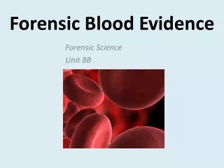
International Journal of Trend in Scientific Research and Development (IJTSRD) Volume: 3 | Issue: 2 | Jan-Feb 2019 Available Online: www.ijtsrd.com e-ISSN: 2456 - 6470 Forensic Existence of Blood as a Dynamic Evidence Sanya Sharma1, Shipra Rohatgi2 1BSc Student, Amity School of Applied Sciences, 2Research Scholar, Amity Institute of Forensic Sciences, Amity University, Manesar, Haryana, India ABSTRACT Blood is one of the chief form of biological fluid which is recovered at the location of crime. This review article focus on blood as a prime forensic evidence which is most commonly encountered on crime scenes. The font of insipiration for the same is the variable facts and the frequent occurrence of blood as an evidence on the crime scene. The article pronounces the complete details of blood, including its composition, cell types, relation of blood to scientific science, handling (liquid blood stains , wet blood stains, dried blood stains, and the post mortum blood) and preservation methods followed by a detailed analysis (preliminary and confirmatory test). The analysis of blood can be categorised on the basis of its physical and chemical properties. The major aim of this study is to provide all the relevant facts related to blood collection, packaging and the entire examination. KEYWORDS: Erythrocytes, Plasma, ABO system, collection and packaging, Blood spatter INTRODUCTION Blood is a complex viscous biological fluid which helps in the transpotation of oxygen and important nutrients to the cells and fading of carbon dioxide and other waste products. Blood contains erythrocytes (RBC), WBC, and Platelets.[3] Blood lies in the category of tissue as well as biological fluid because it is a collection of specialised cells that perform a similar function and a fluid because it contains water in a large amount. Blood consist of 55% of plasma and 45% of cell. Blood is a sticky, opaque fluid with a characteristic metallic taste. As children, we discover its saltiness the first time we stick a cut finger into our mouth. Depending on the amount of oxygen it is carrying, the colour of blood varies from scarlet(oxygen rich) to dark red (oxygen poor).Blood is more dense than water and about five times more viscous, largely because of its formed elements. Blood is slightly alkaline, with pH ranging from 7.35 to 7.45 approximately Table 1: Depicting the Composition of plasma and temperatue of about 38C.Blood accounts for approximately 8% of body weight. Its average volume in healthy adult male is 5-6L (about 1.5gallons), somewhat greater than in healthy adult females (4-5L). [1] These cells are suspended in a liquid matrix called plasma which makes blood a fluid. [2] Plasma is yellowish in colour, it contains proteins (fibrinogen or thrombin and traces of insulin, blinoribin etc.). It contains glucose, dissolved ions and platelets or thrombocytes which that helps in blood clotting, transpotation of enzymes and some important nutrients. It also performs some econdary functions and perform temperature regulation in human body. The different types of blood cells are mjorly produced in the region of bone marrow, located in the thigh bone or femue bone of the human body. Constituents Description and importance 90%of plasma volume; dissolving medium for blood;heat absorbing 8% of plasma volume; contribute to osmotic pressure maintanence. All the proteins are formed in liver. 60% of plasma protein;main contributor to osmotoic pressure. 36% of plasma proteins.most are transport proteins that binds lipids,metal ions and fat-soluble vitamins. 4% of plasma proteins;forms fibrin threads of blood clot. By-products of cellular metabolism,such as urea,uric acid, creatinine and ammonium salts. Materials absorbed from digestive tract and transported for use throughout the body. Water Solutes Plasma proteins a)Albumin b)Globulins Alpha, Beta and Gamma c)Fibrinogen Non protein nitrogenous substances Nutrients (organic) Electrolytes Cations which helps in maintaing the ph of blood. Respiratory gases Oxygen and carbon dioxide. Hormones Steroid and thyroid hormones carried by plasma proteins. @ IJTSRD | Unique Reference Paper ID – IJTSRD21396 | Volume – 3 | Issue – 2 | Jan-Feb 2019 Page: 467
International Journal of Trend in Scientific Research and Development (IJTSRD) @ www.ijtsrd.com eISSN: 2456-6470 Red blood cells(Erythrocytes) The process of formation of RBC is called erythropoiesis. The stem cell is proerythroblasts. It takes place in red bone marrow and minimum time involved for the formation of RBC is 7 days. The rate of erythropoiesis is 2.5 million RBC/second. The growth of RBC starts from the stem cell which is proerythroblasts and the very first phase of the life cycle is early erythroblast in which the nucleus is present and a very distinct and big cell membrane is also present , the second phase is late erythroblast the chromatin condenstation starts from first phase and last till the second phase and the nucleus polarization begins in this phase, the third phase is normoblast in which the contractile actin ring of nucleus is formed, the fourth phase is reticulocyte in which the nucleus is completely discharged out from the cell and finally the erythrocyte is formed as the last phase of the cycle . Concentration in blood is 4-6 million/cubic mm varies with gender differences and high individual variability. Morphology:- biconcave discs,large surface area and enables cells to bend in small capillaries. Main characteristics:- reduced cell, no nucleus(cannot reproduce average life span: 120 days), no mitochondria(no metabolism), no ribosomes( no protein synthesis). Function:- transport haemoglobin (280 million haemoglobin molecules/cell), contain high concentration of carbonic anhydrase, contain high concentration chlorine bicarbonate pump.[5] Fig2:- Structure of hemoglobin White blood cells (Leukocytes) Leukocytes form a protective, movable army that helps the body in defending against damage by bacterial,viral and parasitic attack. Red blood cells are confined to the blood stream and functions in the blood whereas White blood cells, by contrast, are able to slip into and out of blood vessels (by process called diapedesis). The circulatory system is simply their means of transpiration to areas of the body where their services are needed for inflammatory or immune responses. WBCs are classified into two major groupsi.e. granulocytes which contain granules(it is a type of vesical which is filled with fluid or a sac like structure which is filled with fluid) in their cytoplasm and another is agranulocytes which don’t contain granules in their cytoplasm. The granulocytes are further divided into three different cells i.e. Neutrophils they have multi-lobed nucleus, Eosinophils they have kidney or bean shaped nucleus and Basophils they have bilobed nucleus. The stem cell for granulocytes is myeloblasts. The agranulocytes are divided into two different categories lymphocytes they have circular shaped nucleus (stem cell = lymphoblasts) and monocytes they have horse-shoe shaped nucleus (stem cell = monoblasts). [7] Platelets (Thrombocytes) The stem cell of platelets is megakaryoblasts. Platelets are trivial anucleate cell fragments that circulate in blood playing decisive role in managing vascular integrity and regulating hemostatics. Plateltes are also involved in the fundamental biological process of chronic inflammation associated with disease pathology. Platelets indices like mean platelets volume (MPV), platelets distributed width (PWD), and platelets crit (PCT) are useful as cheap noninvasive biomarkers for accessing the diseased states. Dynamic platelets bear distinct morphology, where alpha and dense granule are actively involved in secretion of molecules like GPllb , llla , fibrinogen, Vwf, catecholamines, serotonin, calcium, ATP, ADP, and so forth, which are involved in aggregation. Differential expression of surface receptors like CD36, CD41, CD61 and so forth have also been quantitated in several disease.[8] ABO blood group system Landsteiner established that serum of one individual would clump (agglutinate) the cells of another individual. This is because the cells contain a substance known as an antigen and the serum contains antibodies. Two types of antigens and antibodies are known i.e antigens A and B and antibodies anti-A(alpha) and anti-B(beta). In the blood of a human being, A and B antigens may occur separately, i.e. A Fig1:- Structure of Erythrocyte Hemoglobin A hemoprotein found only in red blood cells. The normal level of haemoglobin in males is 14-16 (g/Dl) and females is 13-15 (g/Dl). Hemoglobin A(HbA) is a major Hb in adults and is composed of four polypeptide chains: two alpha and beta chains. It also contains two dimers of alpha and beta subunits. It contains heme as prosthetic group. Heme reversibly binds to oxygen ( reversible binding is whenever the oxygen is high it takes it and whenever it is low it releases it). It contains protoporphyrine 9 and ferrous iron (fe2+). Fe2+ is present in the center of the heme. Fe2+ binds to four nitrogen atoms of the porphyrin ring. It forms two additional bonds with histidine residue of globin chain and oxygen. The dimers are held together by non-covalent interactions. Each chain is a subunit with a heme group in the center that carries oxygen. A Hb molecule contains four heme groups and carries four molecules of O2. Its fuction is to transport oxygen, carries oxygen from the lungs to tissues, carries carbon dioxide from tissues back to the lungs.[6] @ IJTSRD | Unique Reference Paper ID – IJTSRD21396 | Volume – 3 | Issue – 2 | Jan-Feb 2019 Page: 468
International Journal of Trend in Scientific Research and Development (IJTSRD) @ www.ijtsrd.com eISSN: 2456-6470 alone or B alone or they may be found together i.e. AB, or they may be totally absent,i.e O. A person having A antigen in his red blood cells has group A blood, a person having B antigens has group B blood group, a person having both A and B antigens has group AB blood and a person who has neither A nor B antigens in his blood cells, has group O blood. A person having A antigen in his red blood cells, cannot have an anti-A antibody in his serum, for this would clump his own cells. The same is true of individuals having other antigenic properties. Table No2:- Depicting Blood group percentage in Indian population Antigens in red blood cells A A Anti-B(beta) B B Anti-A(alpha) AB A and B Anti-A(alpha) and anti-B(beta) With specific serum, anti-A and anti-B, it is possible to determine the blood group of any blood in the ABO system accurately. Blood group A will agglutinate by anti-A serum which is the serum of B blood groUP; blood group B will agglutinate by anti-B serum; AB blood will agglutinate by both anti-A and anti-B serum; blood of group O will not agglutinate by either anti-A or anti-B serum. Using anti-A and anti-B serum can directly test fresh liquid blood, but in dried blood stains blood group determination is somewhat more complicated. The absorptionelution and the mixed agglutination tests are most frequently used to determine the blood groups of dried stains.[9] Different blood colors The major type of colour seen in the blood of humans is Red color of blood in humans is because of the red pigment which is haemoglobin. A variety of colors can be seen in the blood due to the presence of some chemicals, pigments etc. Some of the common encountered blood colors are Green color- It is due to a pigment known as biliverdin which is generally present along with the haemoglobin and is known to be produced from the breakdown of haemoglobin in the species of phylum annelida, arthropoda and mollusca. Blue colour- It is due to the presence of hemocyanin. Despite of iron the presence of copper is more pronounced. This color of blood mainly belongs to snails, slugs, clams, octopuses and squids. Yellow colour- This type of blood is a characteristic feature of insects because of the presence of lymph (hemolymph) making it yellow. Orange and violet colour- of the blood is because of the absence of respiratory pigment that is the haemoglobin providing a specific color to the blood cockroaches). [10] Classification of blood stain There are various ways in which blood stains can be classified. The commonly used classification system todat’s era is of S. James, P. Kish and P. Sutton.[11}According to them the blood stains can be divided into three categories Passive blood stains- This category of blood stain patterns are formed under the influence of gravity therefore they are known as passive or gravitational blood stains such type of blood stains are formed as a result of contact between two surfaces among which one should contain the blood. Being the contact stain they provide information about the sequences of movement. Flow patterns, pooling/saturation and drip stains also belong to this category. Aletered blood stains– This area of blood stain pattern involves the blood patterns which are being disturbed from its normal position. Blood Spatter- The third set contains all further stain types, such as blood clots and diluted blood thatbresults from the addition of other liquids.[11] Collection and packaging and preservation of blood or blood stains A.Blood from a person:- it is always necessary to collect reference samples from suspects and victims. In the great majority of cases, these samples consists of liquid blood. Liquid blood from a person should be collected by qualified medical personnel. The crime laboratory should be informed if the subject had recently received a blood transfusion of any kind. Two tubes of blood, about 5ml each, should be collected in vacutainers with EDTA as anticoagulant. In the case of collecting reference samples from post-mortem subjects, a blood sample should be obtained from non body cavity areas such as heart or major internal blood vessels. Each tube should be labelled with the date, time, subject’s name, location, collector’s name, case number and exhibit number. Blood samples must be refrigated, not frozen and submitted to the laboratory as soon as possible.[12] B.Liquid blood specimens at crime scene:- Liquid blood should be collected with a clean (preferable sterile) syring or disposable pipette and transferred to a clean (preferable sterile) test tube. A blood clot can be transferred to a clean test tube with a clean spatula. A clean cotton cloth can be used to soak up liquid blood or a blood clot (avoiding areas containing only serum). Wet blood samples, if they are collected, must be preserved in a suitable anticoagulant and kept in a refrigerator. These specimens should be submitted to the laboratory as soon as possible. Label the specimen with case number, item number, date, time, location and evidence collector’s name.[12] C.Wet bloodstains:- small objects bearing wet bloodstains should be allowed to air dry, then collected as is. An effort should be made to preserve the integrity of any blood stains patterns during packaging and transportation. Large objects that cannot be removed from a crime scene may have wet blood stains on them. The wet blood should be transferred onto clean cotton cloth. Bloodstained cotton cloth must be allowed to air dry befor packaging in a paper container. Each object and container must be properly labelled.[12] D.Dried bloodstains on removable items:- dried blood stains on weapons, garments and other movable objects should be collected separately by collecting the entire item. Each item should be placed in its own (paper) Approx % in Indian population 22 33 5 Blood group Antibodies in serum - - - 40 @ IJTSRD | Unique Reference Paper ID – IJTSRD21396 | Volume – 3 | Issue – 2 | Jan-Feb 2019 Page: 469
International Journal of Trend in Scientific Research and Development (IJTSRD) @ www.ijtsrd.com eISSN: 2456-6470 container, and these should be sealed and labelled properly.[12] E.Dried blood stains on solid, Nonabsorbent surfaces of immovable obejcts:- The blood stain pattern should be documented and sketched to the extent necessary. The stain can be tape lifted or scarped off the object onto a clean piece of paper. The tape lifter or the paper with blood crust can then placed into a “druggist fold”, and placed in an envelope which is sealed. Each item must be labelled properly. If the blood stain cannot be scarped off or the supporter object cannot be cut, then the blood stain may be eluted onto a clean switch, moistened with sterilized saline or water by rubbing the cotton switch on the stained area. The switch is then allowed to dry and is placed in a paper fold packet. The packet is then placed in envelope which is sealed, and properly labelled. Always obtain a control by repeating the procedure on an object but unstained area of the surface containing the blood stain. If the blood stain is located on an object than can be cut, then a portion of item containing the blood stain can be removed by cutting with a clean, sharp instrument. Each cutting should be packaged separately and labelled accordingly. An unstained portion of item should be collected and packaged as a control.[12] Analysis of blood stains A.Physical:- The colour of the blood determines its age if the colour from red changes to maroon this indicates that the blood stain is a day or two older. After that the color becomes dark maroonish and again indicates the blood is old. When the blood is new the stain can easily be collected with swabbing using a cotton sterile cloth but if it is old then it will some what appear like a shrill and cracked sheet which is completely dry. B.Chemical: The chemical analysis of the blood involve preliminary and confirmatory type of examination. 1.Preliminary test- 1.1.Benzidine test- the test is based on the peroxidase acitivity of haemoglobin in blood. A small fibre or scraping is taken on a filter paper and a drop or two of benzidine solution ( 10 percent benzidine in glacial acetic acid) is added to it, followed by hydrogen peroxide. An itense blue colour will develop if blood is present. The test is highly sensitive (1:300,000 dilution of blood), but it is not a specific test for blood, as vegetable peroxidase, chemical oxidants or contamination may give similar reaction. 1.2.Phenolphthalein:- this test is based on the peroxidase acitivity of haemoglobin. Reduced phenolphthalein (colourless) is used as a reagent which add pink color to the sample and presumes the sample as blood. Fig6:- Phenolphthalein test result (Pink Colour) 2.Confirmatory test:- 2.1.Haemin crystal test/ teichman test:- A small crystal of sodium chloride and 2 or 3 drops of glacial acetic acid are placed on a minute fragment of the stain on glass slide. A cover slip is then placed on it and the acid evaporated by heating it gently over a flame. Dark brown rhombic crystals of haemin chloride will be seen under the microscope if the stain is blood. Fig 7 Teichman test (Brown rhombic shaped crystals) 2.2.Takayama test:- The reagents used in this test are saturated solution of glucose, pyridine, 10% of NaOH and distelled test. Haemocromgen crystals are formed which is also called pink needle shaped crystals. It is also performed in the same way as teichman test. Fig 5:- Benzidine test result (Blue colour) Fig 8:- Takayama test (Pink needle shaped crystals) @ IJTSRD | Unique Reference Paper ID – IJTSRD21396 | Volume – 3 | Issue – 2 | Jan-Feb 2019 Page: 470
International Journal of Trend in Scientific Research and Development (IJTSRD) @ www.ijtsrd.com eISSN: 2456-6470 Spectroscopic examination:- the spectroscopic examination is the most delicate, quick and reliable method for detecting haemoglobin. The sample stain is taken in a test-tube. After adding some saline it is examined with a hand spectroscope. Spectral bands characteristics to blood will be observed through the spectroscope.[13] Forensic significance of blood 1.Identification of the biological fluid present at the scene of crime as a blood. 2.Species of origin means that whether the blood is a animal blood or a human blood. 3.The blood which is present at the crime scene belongs to the culprit or the victim. 4.The source estimation of the blood means that whether the blood is menstural, abortion or a vessel flow. 5.The age estimation of the blood. 6.Whether the blood fits from a living or a dead individual. If the blood goes from a living individual then it would be present in the form of scales because of the presence of clotting factor whereas if the blood belongs to the dead individual then it would be in a powder from because of the absence of collting factor. 7.It is mainly used for blood grouping and DNA extraction. Conclusion From the above review article it can be concluded that blood is one of the major and the direct type of biological fluid which is encountered at the crime scene. It is a crucial evidence for thr forensic experts and the further forensic analysis. Blood being the primary type of evidence in almost every type of punishable crimes such as muders, homicides, sexual assaults. The proper collection, packaging and preservation of blood encountered in different forms can be of a great use for the experts to extract the major and the accurate details pertaining to the victim and the culprits. The presence of cells and plasma in the blood can reveal a lot of scientific facts which can help in solving the cases. The major types of analysis performed on the blood which provides accurate and reliable results are the blood grouping and DNA profiling. A series of preliminary and confirmatory test on the blood can narrow down the channel of investigation. Blood helps in the identification of species of origin, the source thst is either from a living or a dead individual, the age of the stain, the genetic makeup of the individual. Sometimes it can also reveal the information about the weapon of offense used by the criminal and the other associative facts that is the presence of any toxicity in the blood. Thertefore the priper handling, documentation followed by in depth the definition of blood is highly important for the success of any case. References [1]https://www.webmd.com> Hear Health [2]Britannica.com/science/blood-biochemistry [3]Elaine N. Marieb, Katija Hoehn, Human anatomy and physiology, 8th ed, Florida, pp635, January 2016. [4]Elaine N. Marieb, Katija Hoehn, Human anatomy and physiology, 8th ed, Florida, pp635, January 2016. [5]Szilvia benko, phd, Red blood cells, Erythrocytes [6]Khaled almohaimede, Abdulaziz Al-shamlan and Moha Khalil, structure and function of haemoglobin. [7]Medical psychology Z.H.AI-Zubaydi [8]Kakali ghosal and Maiture bhattacharya, overview of platelet physiology. [9]B. S. Naber, Forensic science in crime scene investigation, 3rded, Hyderabad, pp118, 2002. [10]Linda crampton, Blood colourin human and animals; meaning and function. [11]SIAK Journal [12]Marian catalin, Anghel Andrei, Oana Mitrasca, Modern methods of collection and preservation of biological evidence for human identification. [13]B. S. Naber, Forensic science in crime scene investigation, 3rded, Hyderabad,pp114-115,2002 @ IJTSRD | Unique Reference Paper ID – IJTSRD21396 | Volume – 3 | Issue – 2 | Jan-Feb 2019 Page: 471