Download

1 / 5
50 likes | 68 Vues
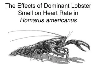
Attempts were made to develop a technique to rear the phyllosoma larvae of Panulirus homarus. The biological characters like fecundity, hatching percentage, larval morphological changes, feed inputs and moulting frequency till the fourth moult were studied. Morphometric and meristic characters of the larvae were also studied till the 42ndday. The larval output was directly proportional to the size of the gravid brood stock. Relationship between the duration of culture X and length of the larvae Y were shown by the relationships Y intercept = 0.5780 u00c2u00b1 0.1074 and X intercept = 0.7283 r2 = 0.8519 . There was significant p 0.0001 positive relationship between total length TL and carapace width CW of phyllosoma larvae. S. Lazarus | J. C. Nisha | R. Thangaraja "Studies on the Phyllosoma Larva of the Indian Rock Lobster, Panulirus Homarus Linnaeus, 1758" Published in International Journal of Trend in Scientific Research and Development (ijtsrd), ISSN: 2456-6470, Volume-4 | Issue-4 , June 2020, URL: https://www.ijtsrd.com/papers/ijtsrd31676.pdf Paper Url :https://www.ijtsrd.com/biological-science/molecular-biology/31676/studies-on-the-phyllosoma-larva-of-the-indian-rock-lobster-panulirus-homarus-linnaeus-1758/s-lazarus<br>

E N D
International Journal of Trend in Scientific Research and Development (IJTSRD) Volume 4 Issue 4, June 2020 Available Online: www.ijtsrd.com e-ISSN: 2456 – 6470 Studies on the Phyllosoma Larva of the Indian Rock Lobster, Panulirus Homarus Linnaeus, 1758 S. Lazarus, J. C. Nisha, R. Thangaraja Institute for Environmental Research and Social Education, Nesamony Nagar, Nagercoil, Tamil Nadu, India ABSTRACT Attempts were made to develop a technique to rear the phyllosoma larvae of Panulirus homarus. The biological characters like fecundity, hatching percentage, larval morphological changes, feed inputs and moulting frequency till the fourth moult were studied. Morphometric and meristic characters of the larvae were also studied till the 42ndday. The larval output was directly proportional to the size of the gravid brood stock. Relationship between the duration of culture (X) and length of the larvae (Y) were shown by the relationships Y intercept = - 0.5780 ± 0.1074 and X intercept = 0.7283 (r2 = 0.8519). There was significant (p<0.0001) positive relationship between total length (TL) and carapace width (CW) of phyllosoma larvae. KEYWORDS: Phyllosoma; spawners; morphometric; meristic; fecundity; moulting How to cite this paper: S. Lazarus | J. C. Nisha | R. Thangaraja "Studies on the Phyllosoma Larva of the Indian Rock Lobster, Panulirus Homarus Linnaeus, 1758" Published in International Journal of Trend in Scientific Research and Development (ijtsrd), ISSN: 2456-6470, Volume-4 | Issue-4, June 2020, pp.1740- 1744, www.ijtsrd.com/papers/ijtsrd31676.pdf Copyright © 2020 by author(s) and International Journal of Trend in Scientific Research and Development Journal. This is an Open Access article distributed under the terms of the Creative Commons Attribution License (CC (http://creativecommons.org/licenses/by /4.0) IJTSRD31676 URL: BY 4.0) INTRODUCTION Aquaculture practice is gaining wide spread importance all over the world as a source of aquatic animal protein. Lobsters, crabs and scallops continue to attract much research attention today because of their demand. The success of aquaculture depends greatly on the development of larval rearing technique. For hatchery production of any commercially important larvae, the stage- wise identification during metamorphosis is very important. The larvae can be fed with a particular feed only after identifying the larval stages (Lazarus et al, 2000). The commercial, recreational and traditional values of spiny lobsters in many countries have led to considerable scientific study, particularly in Panulirus spp. But larval development of Panulirus homarus is still poorly understood. The spiny lobster, Panulirus homarus, is having a very high economic value and demand. But its production through hatcheries is not possible yet because of the poor understanding of the larval development and behaviour. The larvae exhibit a long pelagic and planktonic phase, which makes it difficult to maintain in the hatchery, under controlled conditions. The first success in rearing a palinurid lobster from egg to peurulus stage was achieved by Kittaka (1988), Kittaka and Ikegami, (1988) and Kittaka et al. (1988). Malachite green exposed phyllosoma larvae were examined through six developmental stages by nine moults. Radhakrishnan and Vijayakumaran (1995). Maharajan et al. (2012) studied the fecundity and viability of eggs of spiny lobsters, Panulirus homarus. The survival and growth of phyllosoma larvae of the spiny lobster, Panulirus homarus under different salinity regimes were studied by Dilip et al., (2010). Captive breeding of the spiny lobster, Panulirus homarus was studied by Vijayakumaran et al., (2005). In the present work, an attempt was made to study the fecundity, hatching percentage, larval morphological changes and moulting frequency of the larvae till the fourth moult were made. MATERIAL AND METHODS The exogenous spawners of Panulirus homarus were collected from a landing centre in Muttom, south west coast of India. Three stages of spawners based on the colour of the egg mass such as orange, reddish orange and pale brown were selected as source of spawners for the present study. The disease free and healthy spawners having dark brown eggs were used for spawning. The collected spawners were brought to the hatchery by using a special transportation device fabricated by Babu and Marian (1998) and acclimatized to the hatchery acclimatization, the brooders were dipped in fresh water for 30 seconds to eliminate external fungi and bacteria. After that they were treated with 10 ppm formalin for 2 min. The treated brooders were again washed in fresh sea water to remove the formalin effect found on the exogenous eggs following Babu et al., (2001a). conditions. After @ IJTSRD | Unique Paper ID – IJTSRD31676 | Volume – 4 | Issue – 4 | May-June 2020 Page 1740
International Journal of Trend in Scientific Research and Development (IJTSRD) @ www.ijtsrd.com eISSN: 2456-6470 The brooders of first two maturation stages, with orange and red coloured eggs, were not stocked in the hatching tank. They were maintained in the maturation tank and fed with clams and muscles and water exchange till the egg reached the third stage (i.e.,) the brown coloured egg stage. The 3rd stage exogenous animal with dark brown eggs obtained both from natural source and maturation tanks were transferred in to the hatching tank for the release of young ones from the eggs. In the maturation tank, the brooders were fed with squid and mussel meat at a rate of 20% body weight and 50% water exchange was carried out daily. The stress during this stage was minimized in order to prevent the premature shedding of eggs. The hatching was confirmed in the tanks by the vigorous pleopod movements of the mother lobster and following larval release in the hatching tank. The hatched larvae were separated intermittently from the spawning tank and stocked in larval rearing tanks of 200 L capacity at the rate of 10 nos./L stocking density, where the larvae were fed with Nannochloropsis, an ultra plankton, at the rate of 1 lakh cells/ml concentration. From the second day onwards, the larvae were fed with newly hatched Artemia nauplii and micro algae. The physical parameters maintained during the culture were temperature 30 ± 2° C, salinity 35 ̊/°°, dissolved oxygen 5 mg/l and pH 8.0 ± 2 (Lazarus et al., 2000). Following the method of Babu et al. (2001a), cradle aeration system was improvised for a homogenized and adequate air supply to the rearing system. About 50-90% of sea water exchange was carried out daily. The morphological changes observed in antennules, antenna, head, eye, eyestalk, I maxilliped, II maxilliped, III maxilliped, I, II, III and IV pereiopod, setae, spines and cephalothorax were observed using light microscope and all the morphological variations were drawn using camera lucida. The fecundity, larval moult, growth rate and survival rates were also recorded. The fecundity was recorded in relation with spawner size. The exogenous spawners of a visually acceptable size were allowed to release their eggs as hatched out larvae. After the release of larvae, both released larvae and unhatched eggs were counted. From the counted value, the total fecundity were calculated. Percentage of survival was calculated on the basis of daily mortality. The growth rate was determined microscopically by measuring the total length (TL) and carapace width (CW) of larvae during every moult. Statistical Analysis: Coefficient of regression analysis was performed for total length (CL) and carapace width (CW). Data analysis were based on mean ± SEM. Turkey’s Multiple Comparison Test was performed for moulting frequency and fecundity with 95% confidence level. This statistical analysis was performed by using the software Graph Pad Version 4.0. RESULTS Metamorphosis Before 1st moult In the newly hatched larvae, the head was cone-shaped. The antennules and antennae were uniramous. The eyes were without eyestalk. Mandibles slightly developed. First and second maxillae were seen as rudiments. Second maxilliped was uniramous with 4 segments. First pereiopods were biramous and segmented. Endopod 3 segmented, ends with long narrow spine, surrounded by a group of 7 small spines. The terminal segment was spiny and the 2nd pereiopod rudimentary (Fig. 1). Afer 1st moult Head was slightly narrower at the anterior region and broader at the posterior region. Antennule uniramous, spiny and unsegmented with 3 aesthetases and 2 setae in the terminal region. Antennae uniramous, unsegmented with 2 pairs of setae. Distinct eyestalk was observed. Mandibles slightly developed. 1st and 2nd maxilla were seen rudimentary. 2nd maxilliped uniramous, four segmented. Last segment had 2 setae and third segment had 6 setae. 1st pereiopod biramous, endopod 3 segmented, ends in a long narrow spine, endopod 7 segmented with 6 pairs of plumose setae and the 4th pereiopod seen as rudiment (Fig.2). After 2nd moult Head, narrow in the anterior region and broader at the posterior region. Antennae and antennules uniramous. Distinct eye stalk. 1st and 2nd maxilla rudiments, mandibles slightly developed. 1st maxilliped uniramous, 2 segmented. Second maxilliped uriramous, 5 segmented and segments had 3 setae. Third segment had 6 setae. First pereiopod biramous, exopod 8 segmented with 7 pairs of plumose setae. Towards the end of the stage, 9 segment and 8 pairs of plumose setae were seen. Fourth pereiopod was rudimentary (Fig. 3). After 3rd moult Head, narrower in the anterior region and broader at the posterior region. Antennae and antennules uniramous. Distinct and elongated eye stalk. First and second maxilla and mandibles slightly developed. Second maxilliped uniramous, 5 segmented, last segment had 3 setae and third segment had 6 setae. First pereiopod biramous, exposed, 10 segmented with 9 pairs of plumose setae. Fourth pereiopod slightly developed (Fig.4). After 4th moult There was no change in the shape of cephalic shield, antennae and antennules, distinct and elongated eye stalk. 1st and 2nd maxillae and mandibles slightly developed. First maxillipede 3 segmented, second maxillipede uniramous and 5 segmented, last segment had 3 setae. Third maxillipede bigamous with exopod setae. First and second pereiopods biramous, 7 segmented with 9 pairs of plumose setae in the exopod. Endopod ends with long narrow spine surrounded by a group of small spines. The terminal segment was spiny, and the third periopod was biramous with five pairs of exopod setae. Fourth pereiopod slightly developed and bi- segmented (Fig. 5). Growth rate The growth parameter of the relationship of total length (TL) and carapace width (CW) of phyllosoma larvae is shown in the Fig. 6. Measurement of total length and carapace width of the larvae was carried out up to 42 days. The newly hatched larvae had a total length and carapace width of 1.3280 ± 0.0360 mm and 0.6241± 0.0561mm respectively. On the 11th day, the larval length and width observed were 1.6144 ± 0.1290 mm and 0.7960 ± 0.063mm respectively. On 21st day the measured length and width were 1.7004 ± 0.0993mm and 0.8266 ± 0.0450 mm respectively. On the 31st day the standard length and width of the larvae observed were 2.283 @ IJTSRD | Unique Paper ID – IJTSRD31676 | Volume – 4 | Issue – 4 | May-June 2020 Page 1741
International Journal of Trend in Scientific Research and Development (IJTSRD) @ www.ijtsrd.com eISSN: 2456-6470 ± 0.0116 mm and 1.0346 ± 0.0565 mm respectively and on the 41st day the standard total length and carapace width of the larvae observed were 2.894 ± 0.6210 mm and 2.017 ± 0.1432 mm respectively (Fig. 6). The slope of the regression data showed positive and highly significant (p<0.0001) relationship between total length (TL) and carapace width (CW) of phyllosoma larvae. Fig.4. Phyllosoma larva after 3rd moult At – Antenna, An – Antennule, Max I – Maxillipede I, Max II – Maxillipede II, Max III – Maxillipede III, P1 – Pereiopod I, P2 – Pereiopod II, P3 – Pereiopod III, P4 – Pereiopod IV, Ab – Abdomen, CS – Chephalic shield Fig.1. Newly hatched Phyllosoma larva Fig.5. Phyllosoma larva after 4th moult At – Antenna, An – Antennule, Max I – Maxillipede I, Max II – Maxillipede II, Max III – Maxillipede III, P1 – Pereiopod I, P2 – Pereiopod II, P3 – Pereiopod III, P4 – Pereiopod IV, Ab – Abdomen, CS – Chephalic shield Fig.2. Phyllosoma larva after 1st moult At – Antenna, An – Antennule, Max III – Maxillipede III, P1 – Pereiopod I, P2 – Pereiopod II, P3 – Pereiopod III, P4 – Pereiopod IV, Ab – Abdomen, CS – Chephalic shield, ExB – Exopod budding Fig.6. Percentage of growth rate of phyllosoma larvae p < 0.0001*** (represents highly significance) done by regression analysis Percentage of survival There was not much mortality up to the first 10 days. For the next 10 days, the survival percentage varied between 8.14 and 5.74. After 20 days, the survival percentage started Fig.3. Phyllosoma larva after 2nd moult @ IJTSRD | Unique Paper ID – IJTSRD31676 | Volume – 4 | Issue – 4 | May-June 2020 Page 1742
International Journal of Trend in Scientific Research and Development (IJTSRD) @ www.ijtsrd.com eISSN: 2456-6470 declining from 5.39 to 2.67. From 31st day onwards, the survival percentage further declined and its was 0.01% on the 42nd day (Fig. 7). n=3, p < 0.001** (represents the statistical significance) done by ANOVA, followed by Turkey’s multiple comparison tests DISCUSSION When comparing the present result, with Sankolli and Shenoy (1973) findings, the antennule, antennae, mandible, first maxilla, second maxilla and second maxilliped had similar morphological changes. But the following parts observed by them showed variation with the present work. The first maxilliped was undeveloped, the third maxilliped was uniramous, the exopod of first and second periopod had 8 segments and 7 pairs of plumose setae. But in the present study, the larvae exhibit developed first maxilliped and biramous third maxilliped. In the present case, the exopod of first and second pereiopod had 7 segments and 6 pairs of plumose setae each. Again, the present work showed similar morphological changes in the following parts such as eye, eye stalk, antenna, mandible, first maxilla, second maxilliped, and third maxilliped. They observed four apical setae in the second maxilla, but no such setae were observed in the present study. Nine pairs of setae were recorded by them in the first and second pereiopod, but in the present study only 7 pairs of setae were observed. A study conducted by Radhakrishnan and Vijayakumaran (1993) on phyllosoma larvae of Panulirus homarus showed un segmented eye stalk during the first moult. The present study also confirmed their findings. The carapace width observed by them was 0.72 mm and in the present study it was 0.68 mm. The early stages of the Panulirus echinatus moulted eight times and morphological changes of each appendage were described Abrunhosa et al. (2008). Again a short span study conducted by Radhakrishnan et al., (2009) on the survival and growth of phyllosoma larvae of the tropical spiny lobster, Panulirus homarus maintained with microalgae Nannochloropsis salina culture treatments and without microalgae culture, the author reported that the phyllosoma larvae moulted nine (1- VIII stages) and six times (I – V stages) in the microalgal and non-algal systems, respectively. They found the exopod of fourth pereiopod appeared to become setose in the V stage, the antennule becomes four segmented in the VI stage, 5th periopod appeared as small bud in VII stage, 5th pereiopod budding becomes elongated and formed biramous in stage VIII respectively. At the end, the total length was 5.25 ± 0.04 and carapace width was 3.75 ± 0.04 but at the end of the present study after 4th moult, the total length was 2.89 ± 0.62 mm and carapace width was 2.01 ± 0.14 mm respectively and also showed the entire morphological characters of phyllosoma larvae up to 4th moult. The spawner size also showed direct relationship with nauplii or egg production. The same fact has been observed in P.monodon by (Babu et al., 2001b). About 66% of the breeders belonged to the size group of 61-80 mm CL and this is the dominant size group in the fishery as evident from the export of live lobsters. This size group may be contributing more to the reproduction and recruitment in P. homarus. This may be due to the deposition of more nutritional reserve in the body or its increased age. The fast growth and high survival of early stage phyllosoma larvae of P. homarus in culture system circulated with N. salina and continuous enrichment of Artemia by feeding on N.salina in the micro-algal system was reported by, Fig.7. Percentage of survival of phyllosoma larvae Moulting frequency The first moult was observed after 9.00 ± 0.57 days of hatching. The second, third and fourth moultings were observed on the 19.00 ± 0.57, 28.00 ± 0.57 and 41.33± 0.88 days respectively. There was significant (p < 0.001) difference between the days taken for each moult up to fourth (Fig.8). Fig.8. Moulting frequency of phyllosoma larvae n=3, p < 0.001** (represents the statistical significance) done by ANOVA, followed by Turkey’s multiple comparison tests. Fecundity Number of larvae increased significantly (p < 0.001) with size of the mother lobster (Fig. 9). The spawner of 300gm size yielded 38,046 ± 26.85 larvae and 400 gm size brooder released 45,027 ± 20.51 larvae; the brooder of 550 gm size released 50, 104 ± 57.85 larvae. Fig.9. Relationship between brooder size and larval number @ IJTSRD | Unique Paper ID – IJTSRD31676 | Volume – 4 | Issue – 4 | May-June 2020 Page 1743
International Journal of Trend in Scientific Research and Development (IJTSRD) @ www.ijtsrd.com eISSN: 2456-6470 Radhakrishnan et al. (2009). The present study was done under normal condition. The percentage of survival had been declining from 31st day onwards. It was also noticed that the major cause of the larval mortality was the lack of suitable food for different larval stages (Sarasu and George, 1993; Abrunhosa, 2008). ACKNOWLEDGEMENT The authors are thankful to the University Grants Commission, Government of India for providing necessary funds for operating this project and Manonmaniam Sundaranar University, Tirunelveli, TamilNadu for providing Laboratory facilities. REFERENCES [1]Abrunhosa, F. A., A. P Santiago and J. P. Abrunhosa. 2008. The early phyllosoma stages of spiny lobster Panulirus echinatus Palinuridae) reared in the laboratory. Braz. J. Biol., 68(1): 179-186. egg stage to puerulus. Bull. Scient. Japanese Soc. Fish., 54 (3):413-418. [9]Lazarus, S., S. G. P. Vincent and M. M. Babu. 2000. FAA- Rich Marine Microalgae as energy fuel for the early feeding of phyllosoma larvae of the spiny lobster, Panulirus homarus. In National symposium on phycology in the new millennium, Centre for Advanced studies in Botany, University of Madras, Tamil Nadu, India, p.30. [10]Maharajan, A., M. Vijayakumaran, M., S. Rajalakshmi, P. Jayagopal, M. S. Subramanian and M. C. Remani. 2012. Fecundity and viability of eggs in wild breeders of spiny lobsters, Panulirushomarus (Linnaeus, 1758), 2012. Panulirus versicolor (Latrielle, 1804) and Panulirus ornatus (Fabricius, 1798). J. Mar. Biol. Ass. India, 54:201-209. [11]Radhakrishnan, E. V. and M. Vijayakumaran. 1995. Early larval development of the spiny lobster Panulirus homarus (Linnaeus, 1758). Crustaceana., 68 (2):151- 159. Smith, 1869 (Decapoda: [2]Babu, M. M, and M. P. Marian. 1998. Live transport of gravid Penaeus indicus in Coconut mesocarp dust. Aquacultural Engineering, 18:149-155. [12]Radhakrishnan, E. V., Rekha D. Chakraborty, R. Thangaraja and C. Unnikrishnan, 2009. Effect of Nannochloropsis salina on the survival and growth of phyllosoma of the tropical spiny lobster, Panulirus homarus L. under laboratory conditions. J. Mar. Biol. Ass. India, 51 (1): 52 – 60. [3]Babu, M. M, and M. P. Marian and M. R. Kitto. 2001a. A cradle aeration system for hatching Artemia. Aquacultural Engineering, 24: 85-88. [4]Babu, M. M., S. Lazarus, M. P. Marian and M. R. Kitto, 2001b. Factors determining spawning success in Penaeus monodon, NAGA, The ICLARM, Quarterly (Vol.24) Nos. 1& 2. [13]Radhakrishnan, E. V. and M. Vijayakumaran.1993. Early larval Development of the spiny lobster Panulirus homarus (Linnaeus, 1958) reared in the laboratory. Proceeding of the fourth International Workshop on Lobster Biology and Management. [5]Dilip K., M. Vijaykumaran, T. Senthil Murugan, J. Santhanakumar, T. S. Kumar, N. V. Vinithkumar and R. Kirubagaran. 2010. Survival and growth of early phyllosoma stages of Panulirus homarus under different salinity regimes. J. Mar. Biol. Ass. India, 52(2): 215-218. [14]Sankolli, K. N. and Shenoy. 1973. On the laboratory hatched six phyllosoma stages of Scyllarus sardidus (stimpson). Mar. Biol. Assoc. India; 15(1): 218-226. [15]Sarasu, T. N. and M. J. George. 1993. Larval Biology of spiny lobsters of genus Panulirus CMFRI Spl. Publ., 56:45-47. [6]Kittaka, J., 1988. Culture of the palinurid Jasus lalandii from egg to puerulus. Bull. Japanese Soc. scicnt. Fish., 54(1):87-94 [16]Vijayakumaran, M., T. Senthil Murugan, M. C. Remany, T. Mary Leema, J. Dilip Kumar, J. Santhanakumar, R. Venkatesan, M. Ravindran. 2005. Captive breeding of the spiny lobster, Panulirus homarus. New Zealand Journal of Marine and Freshwater Research, vol.39: 325-334. [7]Kittaka, J. and E. I Kegami, 1988 (cf. a). Culture of the palinurid Palinurus elephas from egg to puerulus. Bull. Japanese Soc. Scient. Fish., 54(7):1149-1154. [8]Kittaka, J., M. Iwai and M. Yoshimijra, 1988 (cf. b). Culture of a hybrid of spiny lobster genus Jasus from @ IJTSRD | Unique Paper ID – IJTSRD31676 | Volume – 4 | Issue – 4 | May-June 2020 Page 1744